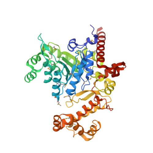
4CW5

Crystal structure of the enoyl reductase domain of DfnA from Bacillus amyloliquefaciens


Experimental Data Snapshot

  • Method: X-RAY DIFFRACTION
  • Resolution: 2.30 Å
  • R-Value Free: 0.231 
  • R-Value Work: 0.204 
  • R-Value Observed: 0.205 

Starting Model: experimental
View more details

wwPDB Validation   3D Report Full Report


Ligand Structure Quality Assessment 


This is version 1.2 of the entry. See complete history


Literature

Evolutionary Origins of the Multienzyme Architecture of Giant Fungal Fatty Acid Synthase.

Bukhari, H.S.Jakob, R.P.Maier, T.

(2014) Structure 22: 1775

  • DOI: https://doi.org/10.1016/j.str.2014.09.016
  • Primary Citation of Related Structures:  
    4CW4, 4CW5

  • PubMed Abstract: 

    Fungal fatty acid synthase (fFAS) is a key paradigm for the evolution of complex multienzymes. Its 48 functional domains are embedded in a matrix of scaffolding elements, which comprises almost 50% of the total sequence and determines the emergent multienzymes properties of fFAS. Catalytic domains of fFAS are derived from monofunctional bacterial enzymes, but the evolutionary origin of the scaffolding elements remains enigmatic. Here, we identify two bacterial protein families of noncanonical fatty acid biosynthesis starter enzymes and trans-acting polyketide enoyl reductases (ERs) as potential ancestors of scaffolding regions in fFAS. The architectures of both protein families are revealed by representative crystal structures of the starter enzyme FabY and DfnA-ER. In both families, a striking structural conservation of insertions to scaffolding elements in fFAS is observed, despite marginal sequence identity. The combined phylogenetic and structural data provide insights into the evolutionary origins of the complex multienzyme architecture of fFAS.


  • Organizational Affiliation

    Biozentrum, Universität Basel, Klingelbergstrasse 50/70, 4056 Basel, Switzerland.


Macromolecules
Find similar proteins by:  (by identity cutoff)  |  3D Structure
Entity ID: 1
MoleculeChains Sequence LengthOrganismDetailsImage
DFNA
A, B
454Bacillus velezensis FZB42Mutation(s): 0 
EC: 2.3.1.39
UniProt
Find proteins for A7Z6E3 (Bacillus velezensis (strain DSM 23117 / BGSC 10A6 / LMG 26770 / FZB42))
Explore A7Z6E3 
Go to UniProtKB:  A7Z6E3
Entity Groups  
Sequence Clusters30% Identity50% Identity70% Identity90% Identity95% Identity100% Identity
UniProt GroupA7Z6E3
Sequence Annotations
Expand
  • Reference Sequence
Small Molecules
Ligands 1 Unique
IDChains Name / Formula / InChI Key2D Diagram3D Interactions
FMN
Query on FMN

Download Ideal Coordinates CCD File 
C [auth A],
D [auth B]
FLAVIN MONONUCLEOTIDE
C17 H21 N4 O9 P
FVTCRASFADXXNN-SCRDCRAPSA-N
Modified Residues  1 Unique
IDChains TypeFormula2D DiagramParent
MSE
Query on MSE
A, B
L-PEPTIDE LINKINGC5 H11 N O2 SeMET
Experimental Data & Validation

Experimental Data

  • Method: X-RAY DIFFRACTION
  • Resolution: 2.30 Å
  • R-Value Free: 0.231 
  • R-Value Work: 0.204 
  • R-Value Observed: 0.205 
  • Space Group: P 21 21 21
Unit Cell:
Length ( Å )Angle ( ˚ )
a = 80.85α = 90
b = 94β = 90
c = 144.53γ = 90
Software Package:
Software NamePurpose
PHENIXrefinement
XDSdata reduction
XDSdata scaling
PHASERphasing

Structure Validation

View Full Validation Report


Ligand Structure Quality Assessment 


Entry History 

Deposition Data

Revision History  (Full details and data files)

  • Version 1.0: 2014-12-17
    Type: Initial release
  • Version 1.1: 2023-12-20
    Changes: Data collection, Database references, Derived calculations, Other, Refinement description
  • Version 1.2: 2024-10-23
    Changes: Structure summary