4CTP | pdb_00004ctp

Structure of rat neuronal nitric oxide synthase heme domain in complex with (S)-6-(2-Amino-2-(3-(2-(6-amino-4-methylpyridin-2-yl) ethyl)phenyl)ethyl)-4-methylpyridin-2-amine


Experimental Data Snapshot

  • Method: X-RAY DIFFRACTION
  • Resolution: 2.05 Å
  • R-Value Free: 
    0.224 (Depositor), 0.224 (DCC) 
  • R-Value Work: 
    0.184 (Depositor), 0.183 (DCC) 
  • R-Value Observed: 
    0.186 (Depositor) 

wwPDB Validation 3D Report Full Report

Validation slider image for 4CTP

Ligand Structure Quality Assessment 


This is version 1.3 of the entry. See complete history

Literature

Nitric Oxide Synthase Inhibitors that Interact with Both a Heme Propionate and Tetrahydrobiopterin Show High Isoform Selectivity.

Kang, S.Tang, W.Li, H.Chreifi, G.Martasek, P.Roman, L.J.Poulos, T.L.Silverman, R.B.

(2014) J Med Chem 57: 4382

  • DOI: https://doi.org/10.1021/jm5004182
  • Primary Citation Related Structures: 
    4CTP, 4CTQ, 4CTR, 4CTT, 4CTU, 4CTV, 4CTW, 4CTX, 4CTY, 4CTZ, 4CU0, 4CU1

  • PubMed Abstract: 

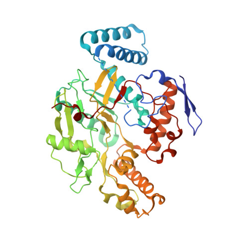
    Overproduction of NO by nNOS is implicated in the pathogenesis of diverse neuronal disorders. Since NO signaling is involved in diverse physiological functions, selective inhibition of nNOS over other isoforms is essential to minimize side effects. A series of α-amino functionalized aminopyridine derivatives (3-8) were designed to probe the structure-activity relationship between ligand, heme propionate, and H4B. Compound 8R was identified as the most potent and selective molecule of this study, exhibiting a Ki of 24 nM for nNOS, with 273-fold and 2822-fold selectivity against iNOS and eNOS, respectively. Although crystal structures of 8R complexed with nNOS and eNOS revealed a similar binding mode, the selectivity stems from the distinct electrostatic environments in two isoforms that result in much lower inhibitor binding free energy in nNOS than in eNOS. These findings provide a basis for further development of simple, but even more selective and potent, nNOS inhibitors.


  • Organizational Affiliation
    • Department of Chemistry, Department of Molecular Biosciences, Chemistry of Life Processes Institute, Center for Molecular Innovation and Drug Discovery, Northwestern University , Evanston, Illinois 60208-3113, United States.

Macromolecule Content 

  • Total Structure Weight: 100.25 kDa 
  • Atom Count: 7,163 
  • Modeled Residue Count: 819 
  • Deposited Residue Count: 844 
  • Unique protein chains: 1

Macromolecules

Find similar proteins by:|  3D Structure
Entity ID: 1
MoleculeChains  Sequence LengthOrganismDetailsImage
NITRIC OXIDE SYNTHASE, BRAIN
A, B
422Rattus norvegicusMutation(s): 0 
EC: 1.14.13.39
UniProt
Find proteins for P29476 (Rattus norvegicus)
Explore P29476 
Go to UniProtKB:  P29476
Entity Groups
Sequence Clusters30% Identity50% Identity70% Identity90% Identity95% Identity100% Identity
UniProt GroupP29476
Sequence Annotations
Expand
Reference Sequence

Small Molecules

Ligands 5 Unique
IDChains Name / Formula / InChI Key2D Diagram3D Interactions
HEM

Query on HEM


Download:Ideal Coordinates CCD File
C [auth A],
H [auth B]
PROTOPORPHYRIN IX CONTAINING FE
C34 H32 Fe N4 O4
KABFMIBPWCXCRK-RGGAHWMASA-L
2IK

Query on 2IK


Download:Ideal Coordinates CCD File
E [auth A],
J [auth B]
6-[(2S)-2-amino-2-{3-[2-(6-amino-4-methylpyridin-2-yl)ethyl]phenyl}ethyl]-4-methylpyridin-2-amine
C22 H27 N5
HPGDXHOKQNTRIC-FQEVSTJZSA-N
H4B

Query on H4B


Download:Ideal Coordinates CCD File
D [auth A],
I [auth B]
5,6,7,8-TETRAHYDROBIOPTERIN
C9 H15 N5 O3
FNKQXYHWGSIFBK-RPDRRWSUSA-N
ZN

Query on ZN


Download:Ideal Coordinates CCD File
G [auth A]ZINC ION
Zn
PTFCDOFLOPIGGS-UHFFFAOYSA-N
ACT

Query on ACT


Download:Ideal Coordinates CCD File
F [auth A],
K [auth B]
ACETATE ION
C2 H3 O2
QTBSBXVTEAMEQO-UHFFFAOYSA-M
Binding Affinity Annotations 
IDSourceBinding Affinity
2IK BindingDB:  4CTP Ki: 144 (nM) from 1 assay(s)

Experimental Data & Validation

Experimental Data

  • Method: X-RAY DIFFRACTION
  • Resolution: 2.05 Å
  • R-Value Free:  0.224 (Depositor), 0.224 (DCC) 
  • R-Value Work:  0.184 (Depositor), 0.183 (DCC) 
  • R-Value Observed: 0.186 (Depositor) 
Space Group: P 21 21 21
Unit Cell:
Length ( Å )Angle ( ˚ )
a = 51.888α = 90
b = 110.994β = 90
c = 164.386γ = 90
Software Package:
Software NamePurpose
REFMACrefinement
HKL-2000data reduction
SCALEPACKdata scaling
REFMACphasing

Structure Validation

View Full Validation Report


Ligand Structure Quality Assessment 


Entry History 

Deposition Data

Revision History  (Full details and data files)

  • Version 1.0: 2014-05-07
    Type: Initial release
  • Version 1.1: 2014-06-04
    Changes: Database references
  • Version 1.2: 2014-08-13
    Changes: Atomic model, Derived calculations, Non-polymer description, Other
  • Version 1.3: 2024-05-08
    Changes: Data collection, Database references, Derived calculations, Other