4COG | pdb_00004cog

Crystal structure of kynurenine formamidase from Burkholderia cenocepacia


Experimental Data Snapshot

  • Method: X-RAY DIFFRACTION
  • Resolution: 1.60 Å
  • R-Value Free: 
    0.184 (Depositor), 0.184 (DCC) 
  • R-Value Work: 
    0.149 (Depositor), 0.148 (DCC) 
  • R-Value Observed: 
    0.151 (Depositor) 

Starting Model: other
View more details

wwPDB Validation 3D Report Full Report

Validation slider image for 4COG

This is version 1.4 of the entry. See complete history

Literature

Structure of Bacterial Kynurenine Formamidase Reveals a Crowded Binuclear-Zinc Catalytic Site Primed to Generate a Potent Nucleophile.

Diaz-Saez, L.Srikannathasan, V.Zoltner, M.Hunter, W.N.

(2014) Biochem J 462: 581

  • DOI: https://doi.org/10.1042/BJ20140511
  • Primary Citation Related Structures: 
    4CO9, 4COB, 4COG, 4CZ1

  • PubMed Abstract: 

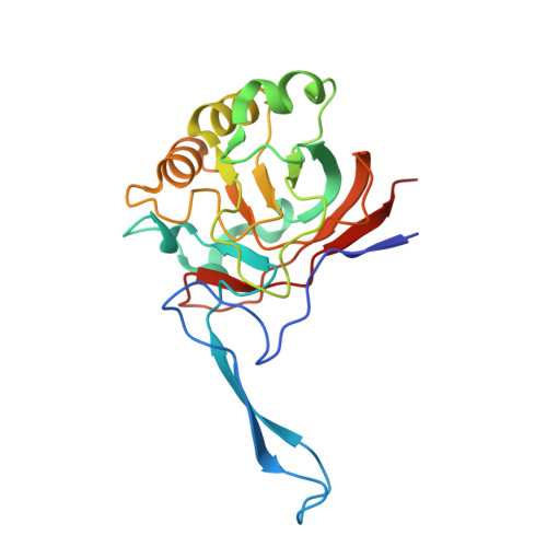
    Tryptophan is an important precursor for chemical entities that ultimately support the biosynthesis of key metabolites. The second stage of tryptophan catabolism is catalysed by kynurenine formamidase, an enzyme that is different between eukaryotes and prokaryotes. In the present study, we characterize the catalytic properties and present the crystal structures of three bacterial kynurenine formamidases. The structures reveal a new amidase protein fold, a highly organized and distinctive binuclear Zn2+ catalytic centre in a confined, hydrophobic and relatively rigid active site. The structure of a complex with 2-aminoacetophenone delineates aspects of molecular recognition extending to the observation that the substrate itself may be conformationally restricted to assist binding in the confined space of the active site and for subsequent processing. The cations occupy a crowded environment, and, unlike most Zn2+-dependent enzymes, there is little scope to increase co-ordination number during catalysis. We propose that the presence of a bridging water/hydroxide ligand in conjunction with the placement of an active site histidine supports a distinctive amidation mechanism.


  • Organizational Affiliation
    • *Division of Biological Chemistry and Drug Discovery, College of Life Sciences, University of Dundee, Dow Street, Dundee DD1 5EH, U.K.

Macromolecule Content 

  • Total Structure Weight: 94.24 kDa 
  • Atom Count: 7,700 
  • Modeled Residue Count: 828 
  • Deposited Residue Count: 860 
  • Unique protein chains: 1

Macromolecules

Find similar proteins by:|  3D Structure
Entity ID: 1
MoleculeChains  Sequence LengthOrganismDetailsImage
KYNURENINE FORMAMIDASE
A, B, C, D
215Burkholderia cenocepacia J2315Mutation(s): 0 
EC: 3.5.1.9
UniProt
Find proteins for B4E9I9 (Burkholderia cenocepacia (strain ATCC BAA-245 / DSM 16553 / LMG 16656 / NCTC 13227 / J2315 / CF5610))
Explore B4E9I9 
Go to UniProtKB:  B4E9I9
Entity Groups
Sequence Clusters30% Identity50% Identity70% Identity90% Identity95% Identity100% Identity
UniProt GroupB4E9I9
Sequence Annotations
Expand
Reference Sequence

Small Molecules

Ligands 6 Unique
IDChains Name / Formula / InChI Key2D Diagram3D Interactions
CD

Query on CD


Download:Ideal Coordinates CCD File
F [auth A],
HA [auth D],
P [auth B],
Z [auth C]
CADMIUM ION
Cd
WLZRMCYVCSSEQC-UHFFFAOYSA-N
PEG

Query on PEG


Download:Ideal Coordinates CCD File
FA [auth C]DI(HYDROXYETHYL)ETHER
C4 H10 O3
MTHSVFCYNBDYFN-UHFFFAOYSA-N
GOL

Query on GOL


Download:Ideal Coordinates CCD File
CA [auth C]
DA [auth C]
EA [auth C]
I [auth A]
J [auth A]
CA [auth C],
DA [auth C],
EA [auth C],
I [auth A],
J [auth A],
K [auth A],
KA [auth D],
LA [auth D],
MA [auth D],
S [auth B],
T [auth B],
U [auth B]
GLYCEROL
C3 H8 O3
PEDCQBHIVMGVHV-UHFFFAOYSA-N
ZN

Query on ZN


Download:Ideal Coordinates CCD File
E [auth A],
GA [auth D],
O [auth B],
Y [auth C]
ZINC ION
Zn
PTFCDOFLOPIGGS-UHFFFAOYSA-N
EDO

Query on EDO


Download:Ideal Coordinates CCD File
L [auth A]
M [auth A]
N [auth A]
NA [auth D]
OA [auth D]
L [auth A],
M [auth A],
N [auth A],
NA [auth D],
OA [auth D],
PA [auth D],
V [auth B],
W [auth B],
X [auth B]
1,2-ETHANEDIOL
C2 H6 O2
LYCAIKOWRPUZTN-UHFFFAOYSA-N
MG

Query on MG


Download:Ideal Coordinates CCD File
AA [auth C]
BA [auth C]
G [auth A]
H [auth A]
IA [auth D]
AA [auth C],
BA [auth C],
G [auth A],
H [auth A],
IA [auth D],
JA [auth D],
Q [auth B],
R [auth B]
MAGNESIUM ION
Mg
JLVVSXFLKOJNIY-UHFFFAOYSA-N

Experimental Data & Validation

Experimental Data

  • Method: X-RAY DIFFRACTION
  • Resolution: 1.60 Å
  • R-Value Free:  0.184 (Depositor), 0.184 (DCC) 
  • R-Value Work:  0.149 (Depositor), 0.148 (DCC) 
  • R-Value Observed: 0.151 (Depositor) 
Space Group: P 1 21 1
Unit Cell:
Length ( Å )Angle ( ˚ )
a = 76.859α = 90
b = 50.12β = 94.15
c = 135.197γ = 90
Software Package:
Software NamePurpose
REFMACrefinement
XDSdata reduction
SCALAdata scaling
MOLREPphasing

Structure Validation

View Full Validation Report


Entry History 

Deposition Data

Revision History  (Full details and data files)

  • Version 1.0: 2014-04-02
    Type: Initial release
  • Version 1.1: 2014-07-02
    Changes: Atomic model, Database references, Derived calculations, Other
  • Version 1.2: 2014-09-03
    Changes: Database references
  • Version 1.3: 2019-03-06
    Changes: Data collection, Experimental preparation, Other
  • Version 1.4: 2024-05-01
    Changes: Data collection, Database references, Derived calculations, Other, Refinement description