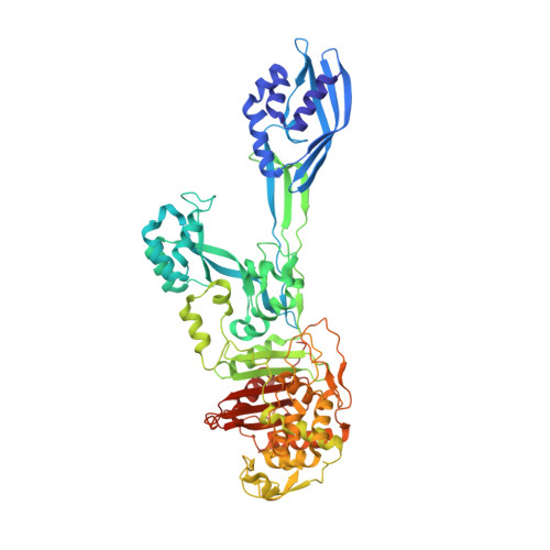
4CJN

Crystal structure of PBP2a from MRSA in complex with quinazolinone ligand


Experimental Data Snapshot

  • Method: X-RAY DIFFRACTION
  • Resolution: 1.95 Å
  • R-Value Free: 0.247 
  • R-Value Work: 0.199 
  • R-Value Observed: 0.201 

Starting Model: experimental
View more details

wwPDB Validation   3D Report Full Report


Ligand Structure Quality Assessment 


This is version 1.3 of the entry. See complete history


Literature

Discovery of Antibiotic (E)-3-(3-Carboxyphenyl)-2-(4-Cyanostyryl)Quinazolin-4(3H)-One.

Bouley, R.Kumarasiri, M.Peng, Z.Otero, L.H.Song, W.Suckow, M.A.Schroeder, V.A.Wolter, W.R.Lastochkin, E.Antunes, N.T.Pi, H.Vakulenko, S.Hermoso, J.A.Chang, M.Mobashery, S.

(2015) J Am Chem Soc 137: 1738

  • DOI: https://doi.org/10.1021/jacs.5b00056
  • Primary Citation of Related Structures:  
    4CJN

  • PubMed Abstract: 

    In the face of the clinical challenge posed by resistant bacteria, the present needs for novel classes of antibiotics are genuine. In silico docking and screening, followed by chemical synthesis of a library of quinazolinones, led to the discovery of (E)-3-(3-carboxyphenyl)-2-(4-cyanostyryl)quinazolin-4(3H)-one (compound 2) as an antibiotic effective in vivo against methicillin-resistant Staphylococcus aureus (MRSA). This antibiotic impairs cell-wall biosynthesis as documented by functional assays, showing binding of 2 to penicillin-binding protein (PBP) 2a. We document that the antibiotic also inhibits PBP1 of S. aureus, indicating a broad targeting of structurally similar PBPs by this antibiotic. This class of antibiotics holds promise in fighting MRSA infections.


  • Organizational Affiliation

    Department of Chemistry and Biochemistry, University of Notre Dame , Notre Dame, Indiana 46556, United States.


Macromolecules
Find similar proteins by:  (by identity cutoff)  |  3D Structure
Entity ID: 1
MoleculeChains Sequence LengthOrganismDetailsImage
PENICILLIN BINDING PROTEIN 2 PRIME
A, B
642Staphylococcus aureus subsp. aureus Mu50Mutation(s): 0 
EC: 3.4.16.4
UniProt
Find proteins for A0A0J9X1X5 (Staphylococcus aureus (strain Mu50 / ATCC 700699))
Explore A0A0J9X1X5 
Go to UniProtKB:  A0A0J9X1X5
Entity Groups  
Sequence Clusters30% Identity50% Identity70% Identity90% Identity95% Identity100% Identity
UniProt GroupA0A0J9X1X5
Sequence Annotations
Expand
  • Reference Sequence
Small Molecules
Ligands 4 Unique
IDChains Name / Formula / InChI Key2D Diagram3D Interactions
QNZ
Query on QNZ

Download Ideal Coordinates CCD File 
L [auth B](E)-3-(2-(4-cyanostyryl)-4-oxoquinazolin-3(4H)-yl)benzoic acid
C24 H15 N3 O3
XWICYONKRAKFAQ-OUKQBFOZSA-N
MUR
Query on MUR

Download Ideal Coordinates CCD File 
G [auth A],
M [auth B]
beta-muramic acid
C9 H17 N O7
MSFSPUZXLOGKHJ-KTZFPWNASA-N
CD
Query on CD

Download Ideal Coordinates CCD File 
C [auth A],
D [auth A],
H [auth B],
I [auth B]
CADMIUM ION
Cd
WLZRMCYVCSSEQC-UHFFFAOYSA-N
CL
Query on CL

Download Ideal Coordinates CCD File 
E [auth A],
F [auth A],
J [auth B],
K [auth B]
CHLORIDE ION
Cl
VEXZGXHMUGYJMC-UHFFFAOYSA-M
Experimental Data & Validation

Experimental Data

  • Method: X-RAY DIFFRACTION
  • Resolution: 1.95 Å
  • R-Value Free: 0.247 
  • R-Value Work: 0.199 
  • R-Value Observed: 0.201 
  • Space Group: P 21 21 21
Unit Cell:
Length ( Å )Angle ( ˚ )
a = 81.05α = 90
b = 103.773β = 90
c = 186.428γ = 90
Software Package:
Software NamePurpose
PHENIXrefinement
XDSdata reduction
SCALAdata scaling
PHASERphasing

Structure Validation

View Full Validation Report


Ligand Structure Quality Assessment 


Entry History 

Deposition Data

Revision History  (Full details and data files)

  • Version 1.0: 2015-02-11
    Type: Initial release
  • Version 1.1: 2015-03-04
    Changes: Database references
  • Version 1.2: 2020-07-29
    Type: Remediation
    Reason: Carbohydrate remediation
    Changes: Data collection, Derived calculations, Other, Structure summary
  • Version 1.3: 2023-12-20
    Changes: Data collection, Database references, Refinement description, Structure summary