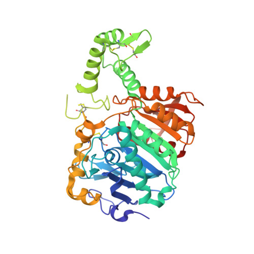
4CIA

Crystal structure of cathepsin A, complexed with compound 1


Experimental Data Snapshot

  • Method: X-RAY DIFFRACTION
  • Resolution: 1.98 Å
  • R-Value Free: 0.232 
  • R-Value Work: 0.170 
  • R-Value Observed: 0.173 

Starting Model: experimental
View more details

wwPDB Validation   3D Report Full Report


Ligand Structure Quality Assessment 


This is version 1.5 of the entry. See complete history


Literature

Crystal structure of cathepsin A, a novel target for the treatment of cardiovascular diseases.

Schreuder, H.A.Liesum, A.Kroll, K.Bohnisch, B.Buning, C.Ruf, S.Sadowski, T.

(2014) Biochem Biophys Res Commun 445: 451-456

  • DOI: https://doi.org/10.1016/j.bbrc.2014.02.014
  • Primary Citation of Related Structures:  
    4CI9, 4CIA, 4CIB

  • PubMed Abstract: 

    The lysosomal serine carboxypeptidase cathepsin A is involved in the breakdown of peptide hormones like endothelin and bradykinin. Recent pharmacological studies with cathepsin A inhibitors in rodents showed a remarkable reduction in cardiac hypertrophy and atrial fibrillation, making cathepsin A a promising target for the treatment of heart failure. Here we describe the crystal structures of activated cathepsin A without inhibitor and with two compounds that mimic the tetrahedral intermediate and the reaction product, respectively. The structure of activated cathepsin A turned out to be very similar to the structure of the inactive precursor. The only difference was the removal of a 40 residue activation domain, partially due to proteolytic removal of the activation peptide, and partially by an order-disorder transition of the peptides flanking the removed activation peptide. The termini of the catalytic core are held together by the Cys253-Cys303 disulfide bond, just before and after the activation domain. One of the compounds we soaked in our crystals reacted covalently with the catalytic Ser150 and formed a tetrahedral intermediate. The other compound got cleaved by the enzyme and a fragment, resembling one of the natural reaction products, was found in the active site. These studies establish cathepsin A as a classical serine proteinase with a well-defined oxyanion hole. The carboxylate group of the cleavage product is bound by a hydrogen-bonding network involving one aspartate and two glutamate side chains. This network can only form if at least half of the carboxylate groups involved are protonated, which explains the acidic pH optimum of the enzyme.


  • Organizational Affiliation

    Sanofi-Aventis Pharma Deutschland GmbH, Industriepark Höchst, 65926 Frankfurt am Main, Germany. Electronic address: herman.schreuder@sanofi.com.


Macromolecules
Find similar proteins by:  (by identity cutoff)  |  3D Structure
Entity ID: 1
MoleculeChains Sequence LengthOrganismDetailsImage
LYSOSOMAL PROTECTIVE PROTEIN455Homo sapiensMutation(s): 0 
EC: 3.4.16.5
UniProt & NIH Common Fund Data Resources
Find proteins for P10619 (Homo sapiens)
Explore P10619 
Go to UniProtKB:  P10619
PHAROS:  P10619
GTEx:  ENSG00000064601 
Entity Groups  
Sequence Clusters30% Identity50% Identity70% Identity90% Identity95% Identity100% Identity
UniProt GroupP10619
Glycosylation
Glycosylation Sites: 2Go to GlyGen: P10619-1
Sequence Annotations
Expand
  • Reference Sequence
Small Molecules
Ligands 3 Unique
IDChains Name / Formula / InChI Key2D Diagram3D Interactions
6KZ
Query on 6KZ

Download Ideal Coordinates CCD File 
B [auth A]N-[(2S)-4-methyl-1-oxidanylidene-1-[[(1R,2S)-1-oxidanyl-1-(5-phenyl-1,2,4-oxadiazol-3-yl)butan-2-yl]amino]pentan-2-yl]morpholine-4-carboxamide
C23 H33 N5 O5
SEDXNAIXMHPLCI-GBESFXJTSA-N
NAG
Query on NAG

Download Ideal Coordinates CCD File 
J [auth A],
K [auth A]
2-acetamido-2-deoxy-beta-D-glucopyranose
C8 H15 N O6
OVRNDRQMDRJTHS-FMDGEEDCSA-N
CD
Query on CD

Download Ideal Coordinates CCD File 
C [auth A]
D [auth A]
E [auth A]
F [auth A]
G [auth A]
C [auth A],
D [auth A],
E [auth A],
F [auth A],
G [auth A],
H [auth A],
I [auth A]
CADMIUM ION
Cd
WLZRMCYVCSSEQC-UHFFFAOYSA-N
Experimental Data & Validation

Experimental Data

  • Method: X-RAY DIFFRACTION
  • Resolution: 1.98 Å
  • R-Value Free: 0.232 
  • R-Value Work: 0.170 
  • R-Value Observed: 0.173 
  • Space Group: C 1 2 1
Unit Cell:
Length ( Å )Angle ( ˚ )
a = 90.106α = 90
b = 101.295β = 101.91
c = 48.077γ = 90
Software Package:
Software NamePurpose
REFMACrefinement
XDSdata reduction
XSCALEdata scaling
PHASERphasing

Structure Validation

View Full Validation Report


Ligand Structure Quality Assessment 


Entry History 

Deposition Data

Revision History  (Full details and data files)

  • Version 1.0: 2014-02-26
    Type: Initial release
  • Version 1.1: 2014-04-02
    Changes: Database references
  • Version 1.2: 2019-03-13
    Changes: Data collection, Database references, Derived calculations, Experimental preparation, Other
  • Version 1.3: 2020-07-29
    Type: Remediation
    Reason: Carbohydrate remediation
    Changes: Data collection, Derived calculations, Other, Structure summary
  • Version 1.4: 2023-12-20
    Changes: Data collection, Database references, Refinement description, Structure summary
  • Version 1.5: 2024-10-16
    Changes: Structure summary