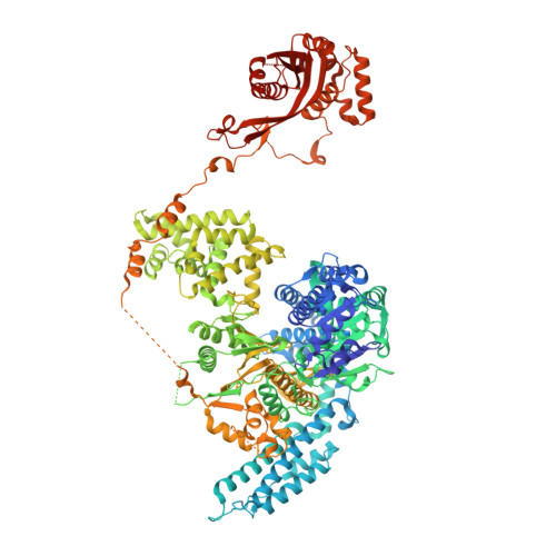
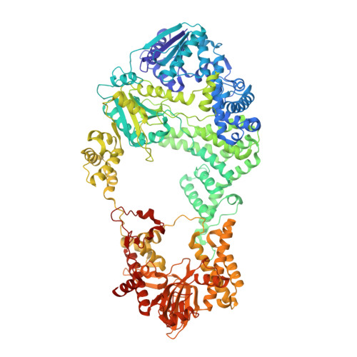
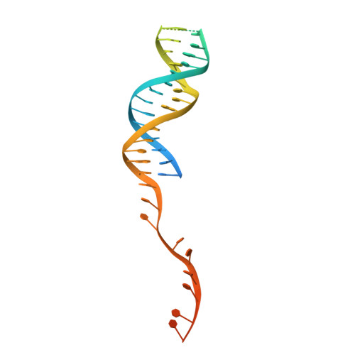
4CEI

Crystal structure of ADPNP-bound AddAB with a forked DNA substrate


Experimental Data Snapshot

  • Method: X-RAY DIFFRACTION
  • Resolution: 2.80 Å
  • R-Value Free: 0.240 
  • R-Value Work: 0.195 
  • R-Value Observed: 0.198 

Starting Model: experimental
View more details

wwPDB Validation   3D Report Full Report


Ligand Structure Quality Assessment 


This is version 1.5 of the entry. See complete history


Literature

Structural Basis for Translocation by Addab Helicase-Nuclease and its Arrest at Chi Sites.

Krajewski, W.W.Fu, X.Wilkinson, M.Cronin, N.B.Dillingham, M.S.Wigley, D.B.

(2014) Nature 508: 416

  • DOI: https://doi.org/10.1038/nature13037
  • Primary Citation of Related Structures:  
    4CEH, 4CEI, 4CEJ

  • PubMed Abstract: 

    In bacterial cells, processing of double-stranded DNA breaks for repair by homologous recombination is dependent upon the recombination hotspot sequence χ (Chi) and is catalysed by either an AddAB- or RecBCD-type helicase-nuclease (reviewed in refs 3, 4). These enzyme complexes unwind and digest the DNA duplex from the broken end until they encounter a χ sequence, whereupon they produce a 3' single-stranded DNA tail onto which they initiate loading of the RecA protein. Consequently, regulation of the AddAB/RecBCD complex by χ is a key control point in DNA repair and other processes involving genetic recombination. Here we report crystal structures of Bacillus subtilis AddAB in complex with different χ-containing DNA substrates either with or without a non-hydrolysable ATP analogue. Comparison of these structures suggests a mechanism for DNA translocation and unwinding, suggests how the enzyme binds specifically to χ sequences, and explains how χ recognition leads to the arrest of AddAB (and RecBCD) translocation that is observed in single-molecule experiments.


  • Organizational Affiliation

    1] Division of Structural Biology, Institute of Cancer Research, Chester Beatty Laboratories, 237 Fulham Road, London SW3 6JB, UK [2] CRT Discovery Laboratories, Department of Biological Sciences, Birkbeck, University of London, London WC1E 7HX, UK.


Macromolecules

Find similar proteins by:  (by identity cutoff)  |  3D Structure
Entity ID: 1
MoleculeChains Sequence LengthOrganismDetailsImage
ATP-DEPENDENT HELICASE/NUCLEASE SUBUNIT A1,232Bacillus subtilis subsp. subtilis str. 168Mutation(s): 2 
EC: 3.1 (PDB Primary Data), 3.6.4.12 (PDB Primary Data)
UniProt
Find proteins for P23478 (Bacillus subtilis (strain 168))
Explore P23478 
Go to UniProtKB:  P23478
Entity Groups  
Sequence Clusters30% Identity50% Identity70% Identity90% Identity95% Identity100% Identity
UniProt GroupP23478
Sequence Annotations
Expand
  • Reference Sequence
Find similar proteins by:  (by identity cutoff)  |  3D Structure
Entity ID: 2
MoleculeChains Sequence LengthOrganismDetailsImage
ATP-DEPENDENT HELICASE/DEOXYRIBONUCLEASE SUBUNIT B1,166Bacillus subtilis subsp. subtilis str. 168Mutation(s): 3 
EC: 3.1 (PDB Primary Data), 3.6.4.12 (PDB Primary Data)
UniProt
Find proteins for P23477 (Bacillus subtilis (strain 168))
Explore P23477 
Go to UniProtKB:  P23477
Entity Groups  
Sequence Clusters30% Identity50% Identity70% Identity90% Identity95% Identity100% Identity
UniProt GroupP23477
Sequence Annotations
Expand
  • Reference Sequence
Find similar nucleic acids by:  (by identity cutoff)  |  3D Structure
Entity ID: 3
MoleculeChains LengthOrganismImage
DNAC [auth X]65synthetic construct
Sequence Annotations
Expand
  • Reference Sequence
Experimental Data & Validation

Experimental Data

  • Method: X-RAY DIFFRACTION
  • Resolution: 2.80 Å
  • R-Value Free: 0.240 
  • R-Value Work: 0.195 
  • R-Value Observed: 0.198 
  • Space Group: P 1 21 1
Unit Cell:
Length ( Å )Angle ( ˚ )
a = 75.944α = 90
b = 151.335β = 95.95
c = 124.56γ = 90
Software Package:
Software NamePurpose
PHENIXrefinement
XDSdata reduction
SCALAdata scaling
PHENIXphasing

Structure Validation

View Full Validation Report


Ligand Structure Quality Assessment 


Entry History 

Deposition Data

Revision History  (Full details and data files)

  • Version 1.0: 2014-03-12
    Type: Initial release
  • Version 1.1: 2014-04-09
    Changes: Database references
  • Version 1.2: 2014-04-23
    Changes: Database references
  • Version 1.3: 2019-01-30
    Changes: Data collection, Experimental preparation, Other
  • Version 1.4: 2019-05-08
    Changes: Data collection, Experimental preparation
  • Version 1.5: 2023-12-20
    Changes: Data collection, Database references, Derived calculations, Other, Refinement description