4CA8 | pdb_00004ca8

Drosophila Angiotensin converting enzyme (AnCE) in complex with a phosphinic tripeptide FII


Experimental Data Snapshot

  • Method: X-RAY DIFFRACTION
  • Resolution: 1.99 Å
  • R-Value Free: 
    0.216 (Depositor), 0.223 (DCC) 
  • R-Value Work: 
    0.194 (Depositor), 0.200 (DCC) 
  • R-Value Observed: 
    0.195 (Depositor) 

Starting Model: experimental
View more details

wwPDB Validation 3D Report Full Report

Validation slider image for 4CA8

Ligand Structure Quality Assessment 


This is version 2.2 of the entry. See complete history

Literature

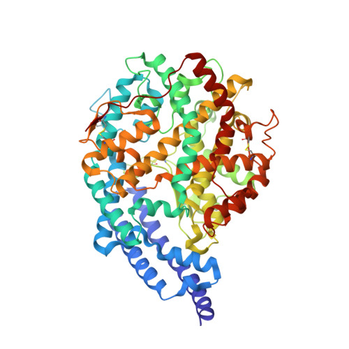
Crystal Structures of Highly Specific Phosphinic Tripeptide Enantiomers in Complex with the Angiotensin-I Converting Enzyme.

Masuyer, G.Akif, M.Czarny, B.Beau, F.Schwager, S.L.Sturrock, E.D.Isaac, R.E.Dive, V.Acharya, K.R.

(2014) FEBS J 281: 943

  • DOI: https://doi.org/10.1111/febs.12660
  • Primary Citation Related Structures: 
    4CA5, 4CA6, 4CA7, 4CA8

  • PubMed Abstract: 

    Human somatic angiotensin-I converting enzyme (ACE) is a zinc-dependent dipeptidyl carboxypeptidase and a central component of the renin angiotensin aldosterone system (RAAS). Its involvement in the modulation of physiological actions of peptide hormones has positioned ACE as an important therapeutic target for the treatment of hypertension and cardiovascular disorders. Here, we report the crystal structures of the two catalytic domains of human ACE (N- and C-) in complex with FI, the S enantiomer of the phosphinic ACE/ECE-1 (endothelin converting enzyme) dual inhibitor FII, to a resolution of 1.91 and 1.85 Å, respectively. In addition, we have determined the structure of AnCE (an ACE homologue from Drosophila melanogaster) in complex with both isomers. The inhibitor FI (S configuration) can adapt to the active site of ACE catalytic domains and shows key differences in its binding mechanism mostly through the reorientation of the isoxazole phenyl side group at the P₁' position compared with FII (R configuration). Differences in binding are also observed between FI and FII in complex with AnCE. Thus, the new structures of the ACE-inhibitor complexes presented here provide useful information for further exploration of ACE inhibitor pharmacophores involving phosphinic peptides and illustrate the role of chirality in enhancing drug specificity.


  • Organizational Affiliation
    • Department of Biology and Biochemistry, University of Bath, UK.

Macromolecule Content 

  • Total Structure Weight: 71.83 kDa 
  • Atom Count: 5,421 
  • Modeled Residue Count: 598 
  • Deposited Residue Count: 598 
  • Unique protein chains: 1

Macromolecules

Find similar proteins by:|  3D Structure
Entity ID: 1
MoleculeChains  Sequence LengthOrganismDetailsImage
ANGIOTENSIN-CONVERTING ENZYME598Drosophila melanogasterMutation(s): 0 
EC: 3.4.15.1
UniProt
Find proteins for Q10714 (Drosophila melanogaster)
Explore Q10714 
Go to UniProtKB:  Q10714
Entity Groups
Sequence Clusters30% Identity50% Identity70% Identity90% Identity95% Identity100% Identity
UniProt GroupQ10714
Glycosylation
Glycosylation Sites: 3Go to GlyGen: Q10714-1
Sequence Annotations
Expand
Reference Sequence

Oligosaccharides

Help  
Entity ID: 2
MoleculeChains Length2D Diagram GlycosylationD Interactions
beta-D-mannopyranose-(1-6)-alpha-D-mannopyranose-(1-3)-[alpha-D-mannopyranose-(1-6)]beta-D-mannopyranose-(1-4)-2-acetamido-2-deoxy-beta-D-glucopyranose-(1-4)-2-acetamido-2-deoxy-beta-D-glucopyranose
B
6N-Glycosylation
Glycosylation Resources
GlyTouCan: G64803HZ
GlyCosmos: G64803HZ
GlyGen: G64803HZ

Small Molecules

Ligands 4 Unique
IDChains Name / Formula / InChI Key2D Diagram3D Interactions
3ES

Query on 3ES


Download:Ideal Coordinates CCD File
C [auth A][(2S)-2-({3-[HYDROXYL(2-PHENYL-(1R)-1-{[(BENZYLOXY)[(2S)-2-({3-[HYDROXYL(2-PHENYL-(1R)-1-CARBONYL]-AMINO}ETHYL)PHOSPHINYL]-2-[(3-PHENYLISOXAZOL-5-YL)METHYL]-1-OXO-PROPYL}AMINO)-3-(4-HYDROXY-PHENYL)
C38 H38 N3 O9 P
ZPFSKFCSVXPMBD-PFESQZPFSA-N
NAG

Query on NAG


Download:Ideal Coordinates CCD File
I [auth A],
J [auth A]
2-acetamido-2-deoxy-beta-D-glucopyranose
C8 H15 N O6
OVRNDRQMDRJTHS-FMDGEEDCSA-N
SO4

Query on SO4


Download:Ideal Coordinates CCD File
D [auth A],
E [auth A],
F [auth A],
G [auth A]
SULFATE ION
O4 S
QAOWNCQODCNURD-UHFFFAOYSA-L
ZN

Query on ZN


Download:Ideal Coordinates CCD File
H [auth A]ZINC ION
Zn
PTFCDOFLOPIGGS-UHFFFAOYSA-N

Experimental Data & Validation

Experimental Data

  • Method: X-RAY DIFFRACTION
  • Resolution: 1.99 Å
  • R-Value Free:  0.216 (Depositor), 0.223 (DCC) 
  • R-Value Work:  0.194 (Depositor), 0.200 (DCC) 
  • R-Value Observed: 0.195 (Depositor) 
Space Group: H 3
Unit Cell:
Length ( Å )Angle ( ˚ )
a = 172.89α = 90
b = 172.89β = 90
c = 100.48γ = 120
Software Package:
Software NamePurpose
REFMACrefinement
MOSFLMdata reduction
SCALAdata scaling
PHASERphasing

Structure Validation

View Full Validation Report


Ligand Structure Quality Assessment 


Entry History 

Deposition Data

Revision History  (Full details and data files)

  • Version 1.0: 2013-12-11
    Type: Initial release
  • Version 1.1: 2014-02-12
    Changes: Database references
  • Version 2.0: 2020-07-29
    Type: Remediation
    Reason: Carbohydrate remediation
    Changes: Atomic model, Data collection, Derived calculations, Other, Structure summary
  • Version 2.1: 2023-12-20
    Changes: Data collection, Database references, Refinement description, Structure summary
  • Version 2.2: 2024-10-09
    Changes: Structure summary