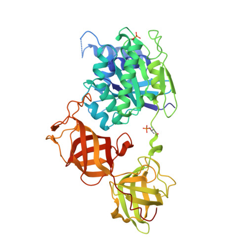
4C0S

Mammalian translation elongation factor eEF1A2


Experimental Data Snapshot

  • Method: X-RAY DIFFRACTION
  • Resolution: 2.70 Å
  • R-Value Free: 0.256 
  • R-Value Work: 0.202 
  • R-Value Observed: 0.205 

wwPDB Validation   3D Report Full Report


Ligand Structure Quality Assessment 


This is version 1.4 of the entry. See complete history


Literature

Mammalian Translation Elongation Factor Eef1A2: X-Ray Structure and New Features of Gdp/GTP Exchange Mechanism in Higher Eukaryotes

Crepin, T.Mccarthy, A.Negrutskii, A.Shalak, V.F.Tukalo, V.Vlasenko, D.O.Yaremchuk, A.D.

(2014) Nucleic Acids Res 42: 12939

  • DOI: https://doi.org/10.1093/nar/gku974
  • Primary Citation of Related Structures:  
    4C0S

  • PubMed Abstract: 

    Eukaryotic elongation factor eEF1A transits between the GTP- and GDP-bound conformations during the ribosomal polypeptide chain elongation. eEF1A*GTP establishes a complex with the aminoacyl-tRNA in the A site of the 80S ribosome. Correct codon-anticodon recognition triggers GTP hydrolysis, with subsequent dissociation of eEF1A*GDP from the ribosome. The structures of both the 'GTP'- and 'GDP'-bound conformations of eEF1A are unknown. Thus, the eEF1A-related ribosomal mechanisms were anticipated only by analogy with the bacterial homolog EF-Tu. Here, we report the first crystal structure of the mammalian eEF1A2*GDP complex which indicates major differences in the organization of the nucleotide-binding domain and intramolecular movements of eEF1A compared to EF-Tu. Our results explain the nucleotide exchange mechanism in the mammalian eEF1A and suggest that the first step of eEF1A*GDP dissociation from the 80S ribosome is the rotation of the nucleotide-binding domain observed after GTP hydrolysis.


  • Organizational Affiliation

    University of Grenoble Alpes, UVHCI, F-38000 Grenoble, France CNRS, UVHCI, F-38000 Grenoble, France Unit for Virus Host-Cell Interactions, University of Grenoble Alpes-EMBL-CNRS, 71 avenue des Martyrs, 38042 France.


Macromolecules
Find similar proteins by:  (by identity cutoff)  |  3D Structure
Entity ID: 1
MoleculeChains Sequence LengthOrganismDetailsImage
ELONGATION FACTOR 1-ALPHA 2
A, B
463Oryctolagus cuniculusMutation(s): 0 
UniProt
Find proteins for Q71V39 (Oryctolagus cuniculus)
Explore Q71V39 
Go to UniProtKB:  Q71V39
Entity Groups  
Sequence Clusters30% Identity50% Identity70% Identity90% Identity95% Identity100% Identity
UniProt GroupQ71V39
Sequence Annotations
Expand
  • Reference Sequence
Experimental Data & Validation

Experimental Data

  • Method: X-RAY DIFFRACTION
  • Resolution: 2.70 Å
  • R-Value Free: 0.256 
  • R-Value Work: 0.202 
  • R-Value Observed: 0.205 
  • Space Group: P 61 2 2
Unit Cell:
Length ( Å )Angle ( ˚ )
a = 135.37α = 90
b = 135.37β = 90
c = 304.62γ = 120
Software Package:
Software NamePurpose
REFMACrefinement
XDSdata reduction
XSCALEdata scaling

Structure Validation

View Full Validation Report


Ligand Structure Quality Assessment 


Entry History 

Deposition Data

Revision History  (Full details and data files)

  • Version 1.0: 2014-08-27
    Type: Initial release
  • Version 1.1: 2014-10-29
    Changes: Database references
  • Version 1.2: 2014-11-26
    Changes: Database references
  • Version 1.3: 2017-01-25
    Changes: Data collection
  • Version 1.4: 2024-11-20
    Changes: Data collection, Database references, Derived calculations, Other, Structure summary