4BX7 | pdb_00004bx7

trans-divalent streptavidin bound to biotin-4-fluorescein


Experimental Data Snapshot

  • Method: X-RAY DIFFRACTION
  • Resolution: 2.26 Å
  • R-Value Free: 
    0.237 (Depositor), 0.245 (DCC) 
  • R-Value Work: 
    0.184 (Depositor), 0.192 (DCC) 
  • R-Value Observed: 
    0.186 (Depositor) 

Starting Model: experimental
View more details

wwPDB Validation 3D Report Full Report

Validation slider image for 4BX7

Ligand Structure Quality Assessment 


This is version 1.5 of the entry. See complete history

Literature

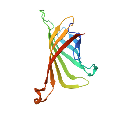
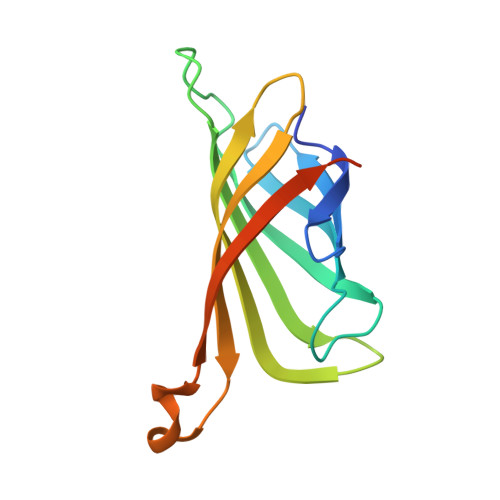
Plug-and-Play Pairing Via Defined Divalent Streptavidins.

Fairhead, M.Krndija, D.Lowe, E.D.Howarth, M.

(2014) J Mol Biology 426: 199

  • DOI: https://doi.org/10.1016/j.jmb.2013.09.016
  • Primary Citation Related Structures: 
    4BX5, 4BX6, 4BX7

  • PubMed Abstract: 

    Streptavidin is one of the most important hubs for molecular biology, either multimerizing biomolecules, bridging one molecule to another, or anchoring to a biotinylated surface/nanoparticle. Streptavidin has the advantage of rapid ultra-stable binding to biotin. However, the ability of streptavidin to bind four biotinylated molecules in a heterogeneous manner is often limiting. Here, we present an efficient approach to isolate streptavidin tetramers with two biotin-binding sites in a precise arrangement, cis or trans. We genetically modified specific subunits with negatively charged tags, refolded a mixture of monomers, and used ion-exchange chromatography to resolve tetramers according to the number and orientation of tags. We solved the crystal structures of cis-divalent streptavidin to 1.4Å resolution and trans-divalent streptavidin to 1.6Å resolution, validating the isolation strategy and explaining the behavior of the Dead streptavidin variant. cis- and trans-divalent streptavidins retained tetravalent streptavidin's high thermostability and low off-rate. These defined divalent streptavidins enabled us to uncover how streptavidin binding depends on the nature of the biotin ligand. Biotinylated DNA showed strong negative cooperativity of binding to cis-divalent but not trans-divalent streptavidin. A small biotinylated protein bound readily to cis and trans binding sites. We also solved the structure of trans-divalent streptavidin bound to biotin-4-fluorescein, showing how one ligand obstructs binding to an adjacent biotin-binding site. Using a hexaglutamate tag proved a more powerful way to isolate monovalent streptavidin, for ultra-stable labeling without undesired clustering. These forms of streptavidin allow this key hub to be used with a new level of precision, for homogeneous molecular assembly.


  • Organizational Affiliation
    • Department of Biochemistry, University of Oxford, South Parks Road, Oxford OX1 3QU, UK.

Macromolecule Content 

  • Total Structure Weight: 28.14 kDa 
  • Atom Count: 1,899 
  • Modeled Residue Count: 237 
  • Deposited Residue Count: 261 
  • Unique protein chains: 2

Macromolecules

Find similar proteins by:|  3D Structure
Entity ID: 1
MoleculeChains  Sequence LengthOrganismDetailsImage
STREPTAVIDIN128Streptomyces avidiniiMutation(s): 3 
UniProt
Find proteins for P22629 (Streptomyces avidinii)
Explore P22629 
Go to UniProtKB:  P22629
Entity Groups
Sequence Clusters30% Identity50% Identity70% Identity90% Identity95% Identity100% Identity
UniProt GroupP22629
Sequence Annotations
Expand
Reference Sequence
Find similar proteins by:|  3D Structure
Entity ID: 2
MoleculeChains  Sequence LengthOrganismDetailsImage
STREPTAVIDIN133Streptomyces avidiniiMutation(s): 0 
UniProt
Find proteins for P22629 (Streptomyces avidinii)
Explore P22629 
Go to UniProtKB:  P22629
Entity Groups
Sequence Clusters30% Identity50% Identity70% Identity90% Identity95% Identity100% Identity
UniProt GroupP22629
Sequence Annotations
Expand
Reference Sequence

Small Molecules

Ligands 2 Unique
IDChains Name / Formula / InChI Key2D Diagram3D Interactions
B4F

Query on B4F


Download:Ideal Coordinates CCD File
D [auth B]biotin-4-fluorescein
C33 H32 N4 O8 S
DFUFXKZUEOKPSD-LFERIPGTSA-N
MPD

Query on MPD


Download:Ideal Coordinates CCD File
C [auth A](4S)-2-METHYL-2,4-PENTANEDIOL
C6 H14 O2
SVTBMSDMJJWYQN-YFKPBYRVSA-N

Experimental Data & Validation

Experimental Data

  • Method: X-RAY DIFFRACTION
  • Resolution: 2.26 Å
  • R-Value Free:  0.237 (Depositor), 0.245 (DCC) 
  • R-Value Work:  0.184 (Depositor), 0.192 (DCC) 
  • R-Value Observed: 0.186 (Depositor) 
Space Group: P 31 2 1
Unit Cell:
Length ( Å )Angle ( ˚ )
a = 64.41α = 90
b = 64.41β = 90
c = 103.13γ = 120
Software Package:
Software NamePurpose
PHENIXrefinement
XDSdata reduction
XSCALEdata scaling
PHASERphasing

Structure Validation

View Full Validation Report


Ligand Structure Quality Assessment 


Entry History 

Deposition Data

Revision History  (Full details and data files)

  • Version 1.0: 2013-09-25
    Type: Initial release
  • Version 1.1: 2013-10-09
    Changes: Database references
  • Version 1.2: 2014-01-15
    Changes: Database references
  • Version 1.3: 2019-03-06
    Changes: Data collection, Experimental preparation, Other
  • Version 1.4: 2019-05-15
    Changes: Data collection, Experimental preparation
  • Version 1.5: 2023-12-20
    Changes: Data collection, Database references, Derived calculations, Other, Refinement description