4BWI | pdb_00004bwi

Structure of the phytochrome Cph2 from Synechocystis sp. PCC6803


Experimental Data Snapshot

  • Method: X-RAY DIFFRACTION
  • Resolution: 2.60 Å
  • R-Value Free: 
    0.253 (Depositor), 0.255 (DCC) 
  • R-Value Work: 
    0.200 (Depositor), 0.205 (DCC) 
  • R-Value Observed: 
    0.203 (Depositor) 

wwPDB Validation 3D Report Full Report

Validation slider image for 4BWI

Ligand Structure Quality Assessment 


This is version 1.5 of the entry. See complete history

Literature

Structure of the Cyanobacterial Phytochrome 2 Photosensor Implies a Tryptophan Switch for Phytochrome Signaling.

Anders, K.Daminelli-Widany, G.Mroginski, M.A.von Stetten, D.Essen, L.-O.

(2013) J Biological Chem 288: 35714

  • DOI: https://doi.org/10.1074/jbc.M113.510461
  • Primary Citation Related Structures: 
    4BWI

  • PubMed Abstract: 

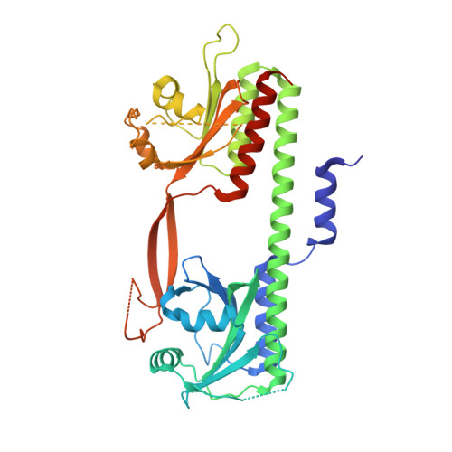
    Phytochromes are highly versatile photoreceptors, which occur ubiquitously in plants as well as in many light-responsive microorganisms. Here, photosynthetic cyanobacteria utilize up to three different phytochrome architectures, where only the plant-like and the single-domain cyanobacteriochromes are structurally characterized so far. Cph2 represents a third group in Synechocystis species and affects their capability of phototaxis by controlling c-di-GMP synthesis and degradation. The 2.6-Å crystal structure of its red/far-red responsive photosensory module in the Pr state reveals a tandem-GAF bidomain that lacks the figure-of-eight knot of the plant/cph1 subfamily. Its covalently attached phycocyanobilin chromophore adopts a highly tilted ZZZssa conformation with a novel set of interactions between its propionates and the GAF1 domain. The tongue-like protrusion from the GAF2 domain interacts with the GAF1-bound chromophore via its conserved PRXSF, WXE, and W(G/A)G motifs. Mutagenesis showed that the integrity of the tongue is indispensable for Pr → Pfr photoconversion and involves a swap of the motifs' tryptophans within the tongue-GAF1 interface. This "Trp switch" is supposed to be a crucial element for the photochromicity of all multidomain phytochromes.


  • Organizational Affiliation
    • From the Department of Chemistry, Biomedical Research Centre, Philipps-Universität, D-35032 Marburg, Germany.

Macromolecule Content 

  • Total Structure Weight: 100.28 kDa 
  • Atom Count: 6,311 
  • Modeled Residue Count: 758 
  • Deposited Residue Count: 864 
  • Unique protein chains: 1

Macromolecules

Find similar proteins by:|  3D Structure
Entity ID: 1
MoleculeChains  Sequence LengthOrganismDetailsImage
PHYTOCHROME-LIKE PROTEIN CPH2
A, B
432Synechocystis sp.Mutation(s): 0 
UniProt
Find proteins for Q55434 (Synechocystis sp. (strain ATCC 27184 / PCC 6803 / Kazusa))
Explore Q55434 
Go to UniProtKB:  Q55434
Entity Groups
Sequence Clusters30% Identity50% Identity70% Identity90% Identity95% Identity100% Identity
UniProt GroupQ55434
Sequence Annotations
Expand
Reference Sequence

Small Molecules

Ligands 4 Unique
IDChains Name / Formula / InChI Key2D Diagram3D Interactions
CYC

Query on CYC


Download:Ideal Coordinates CCD File
D [auth A],
I [auth B]
PHYCOCYANOBILIN
C33 H40 N4 O6
VXTXPYZGDQPMHK-GMXXPEQVSA-N
GLU

Query on GLU


Download:Ideal Coordinates CCD File
C [auth A]GLUTAMIC ACID
C5 H9 N O4
WHUUTDBJXJRKMK-VKHMYHEASA-N
GOL

Query on GOL


Download:Ideal Coordinates CCD File
E [auth A],
J [auth B]
GLYCEROL
C3 H8 O3
PEDCQBHIVMGVHV-UHFFFAOYSA-N
FMT

Query on FMT


Download:Ideal Coordinates CCD File
F [auth A]
G [auth A]
H [auth A]
K [auth B]
L [auth B]
F [auth A],
G [auth A],
H [auth A],
K [auth B],
L [auth B],
M [auth B],
N [auth B]
FORMIC ACID
C H2 O2
BDAGIHXWWSANSR-UHFFFAOYSA-N

Experimental Data & Validation

Experimental Data

  • Method: X-RAY DIFFRACTION
  • Resolution: 2.60 Å
  • R-Value Free:  0.253 (Depositor), 0.255 (DCC) 
  • R-Value Work:  0.200 (Depositor), 0.205 (DCC) 
  • R-Value Observed: 0.203 (Depositor) 
Space Group: P 1 21 1
Unit Cell:
Length ( Å )Angle ( ˚ )
a = 77.43α = 90
b = 88.46β = 109.85
c = 84.87γ = 90
Software Package:
Software NamePurpose
PHENIXrefinement
XDSdata reduction
XSCALEdata scaling
PHENIXphasing

Structure Validation

View Full Validation Report


Ligand Structure Quality Assessment 


Entry History 

Deposition Data

Revision History  (Full details and data files)

  • Version 1.0: 2013-10-30
    Type: Initial release
  • Version 1.1: 2013-11-13
    Changes: Database references
  • Version 1.2: 2014-01-08
    Changes: Database references
  • Version 1.3: 2017-12-13
    Changes: Database references, Structure summary
  • Version 1.4: 2019-05-08
    Changes: Data collection, Derived calculations, Experimental preparation
  • Version 1.5: 2024-11-13
    Changes: Data collection, Database references, Derived calculations, Other, Structure summary