4BUT

Crystal structure of human tankyrase 2 in complex with 4-(4-oxo-3,4- dihydroquinazolin-2-yl)benzene-1-sulfonamide


Experimental Data Snapshot

  • Method: X-RAY DIFFRACTION
  • Resolution: 1.90 Å
  • R-Value Free: 
    0.207 (Depositor), 0.210 (DCC) 
  • R-Value Work: 
    0.171 (Depositor), 0.170 (DCC) 
  • R-Value Observed: 
    0.172 (Depositor) 

wwPDB Validation   3D Report Full Report


Ligand Structure Quality Assessment 

Created with Raphaël 2.3.0Worse 01 BetterLigand structure goodness of fit to experimental dataBest fitted 31FClick on this verticalbar to view details

This is version 1.5 of the entry. See complete history


Literature

Para-Substituted 2-Phenyl-3,4-Dihydroquinazolin-4-Ones as Potent and Selective Tankyrase Inhibitors.

Haikarainen, T.Koivunen, J.Narwal, M.Venkannagari, H.Obaji, E.Joensuu, P.Pihlajaniemi, T.Lehtio, L.

(2013) ChemMedChem 8: 1978

  • DOI: https://doi.org/10.1002/cmdc.201300337
  • Primary Citation of Related Structures:  
    4BU3, 4BU5, 4BU6, 4BU7, 4BU8, 4BU9, 4BUA, 4BUD, 4BUE, 4BUF, 4BUI, 4BUS, 4BUT, 4BUU, 4BUV, 4BUW, 4BUX, 4BUY

  • PubMed Abstract: 

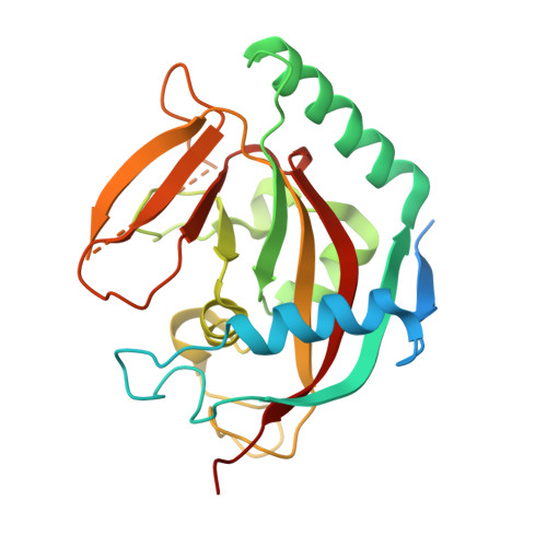
    Human tankyrases are attractive drug targets, especially for the treatment of cancer. We identified a set of highly potent tankyrase inhibitors based on a 2-phenyl-3,4-dihydroquinazolin-4-one scaffold. Substitutions at the para position of the scaffold's phenyl group were evaluated as a strategy to increase potency and improve selectivity. The best compounds displayed single-digit nanomolar potencies, and profiling against several human diphtheria-toxin-like ADP-ribosyltransferases revealed that a subset of these compounds are highly selective tankyrase inhibitors. The compounds also effectively inhibit Wnt signaling in HEK293 cells. The binding mode of all inhibitors was studied by protein X-ray crystallography. This allowed us to establish a structural basis for the development of highly potent and selective tankyrase inhibitors based on the 2-phenyl-3,4-dihydroquinazolin-4-one scaffold and outline a rational approach to the modification of other inhibitor scaffolds that bind to the nicotinamide site of the catalytic domain.


  • Organizational Affiliation

    Department of Biochemistry and Biocenter Oulu, 90014 University of Oulu, PO Box 3000 Oulu (Finland).


Macromolecules
Find similar proteins by:  (by identity cutoff)  |  3D Structure
Entity ID: 1
MoleculeChains Sequence LengthOrganismDetailsImage
TANKYRASE-2
A, B
240Homo sapiensMutation(s): 0 
EC: 2.4.2.30 (PDB Primary Data), 2.4.2 (UniProt)
UniProt & NIH Common Fund Data Resources
Find proteins for Q9H2K2 (Homo sapiens)
Explore Q9H2K2 
Go to UniProtKB:  Q9H2K2
PHAROS:  Q9H2K2
GTEx:  ENSG00000107854 
Entity Groups  
Sequence Clusters30% Identity50% Identity70% Identity90% Identity95% Identity100% Identity
UniProt GroupQ9H2K2
Sequence Annotations
Expand
  • Reference Sequence
Small Molecules
Ligands 5 Unique
IDChains Name / Formula / InChI Key2D Diagram3D Interactions
31F
Query on 31F

Download Ideal Coordinates CCD File 
F [auth A],
L [auth B]
4-(4-oxidanylidene-3H-quinazolin-2-yl)benzenesulfonamide
C14 H11 N3 O3 S
OMUIDCNIRWVPSK-UHFFFAOYSA-N
PEG
Query on PEG

Download Ideal Coordinates CCD File 
H [auth A],
N [auth B]
DI(HYDROXYETHYL)ETHER
C4 H10 O3
MTHSVFCYNBDYFN-UHFFFAOYSA-N
SO4
Query on SO4

Download Ideal Coordinates CCD File 
D [auth A],
E [auth A],
J [auth B],
K [auth B]
SULFATE ION
O4 S
QAOWNCQODCNURD-UHFFFAOYSA-L
GOL
Query on GOL

Download Ideal Coordinates CCD File 
G [auth A],
M [auth B]
GLYCEROL
C3 H8 O3
PEDCQBHIVMGVHV-UHFFFAOYSA-N
ZN
Query on ZN

Download Ideal Coordinates CCD File 
C [auth A],
I [auth B]
ZINC ION
Zn
PTFCDOFLOPIGGS-UHFFFAOYSA-N
Binding Affinity Annotations 
IDSourceBinding Affinity
31F BindingDB:  4BUT IC50: 12 (nM) from 1 assay(s)
Experimental Data & Validation

Experimental Data

  • Method: X-RAY DIFFRACTION
  • Resolution: 1.90 Å
  • R-Value Free:  0.207 (Depositor), 0.210 (DCC) 
  • R-Value Work:  0.171 (Depositor), 0.170 (DCC) 
  • R-Value Observed: 0.172 (Depositor) 
Space Group: C 2 2 21
Unit Cell:
Length ( Å )Angle ( ˚ )
a = 91.5α = 90
b = 97.95β = 90
c = 119.19γ = 90
Software Package:
Software NamePurpose
REFMACrefinement
XDSdata reduction
XSCALEdata scaling

Structure Validation

View Full Validation Report


Ligand Structure Quality Assessment 

Created with Raphaël 2.3.0Worse 01 BetterLigand structure goodness of fit to experimental dataBest fitted 31FClick on this verticalbar to view details

Entry History 

Deposition Data

Revision History  (Full details and data files)

  • Version 1.0: 2013-10-30
    Type: Initial release
  • Version 1.1: 2013-12-11
    Changes: Atomic model
  • Version 1.2: 2018-01-17
    Changes: Data collection
  • Version 1.3: 2019-01-30
    Changes: Data collection, Experimental preparation
  • Version 1.4: 2019-02-06
    Changes: Data collection, Experimental preparation
  • Version 1.5: 2024-05-08
    Changes: Data collection, Database references, Derived calculations, Other