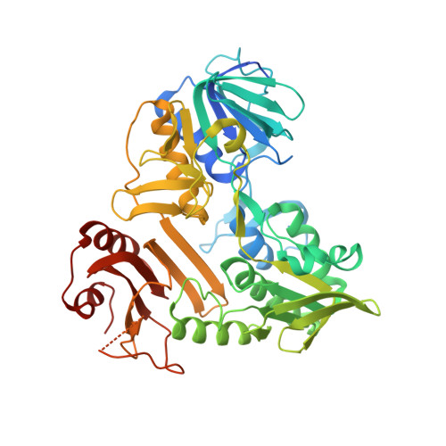
4BUR

Crystal structure of the reduced human Apoptosis inducing factor complexed with NAD


Experimental Data Snapshot

  • Method: X-RAY DIFFRACTION
  • Resolution: 2.88 Å
  • R-Value Free: 0.230 
  • R-Value Work: 0.176 
  • R-Value Observed: 0.180 

Starting Model: experimental
View more details

wwPDB Validation   3D Report Full Report


Ligand Structure Quality Assessment 


This is version 1.2 of the entry. See complete history


Literature

Structural Insights Into the Coenzyme Mediated Monomer-Dimer Transition of the Pro-Apoptotic Apoptosis Inducing Factor.

Ferreira, P.Villanueva, R.Martinez-Julvez, M.Herguedas, B.Marcuello, C.Fernandez-Silva, P.Cabon, L.Hermoso, J.A.Lostao, A.Susin, S.A.Medina, M.

(2014) Biochemistry 53: 4204

  • DOI: https://doi.org/10.1021/bi500343r
  • Primary Citation of Related Structures:  
    4BUR, 4BV6

  • PubMed Abstract: 

    The apoptosis-inducing factor (AIF) is a mitochondrial-flavoprotein that, after cell death induction, is distributed to the nucleus to mediate chromatinolysis. In mitochondria, AIF is present in a monomer-dimer equilibrium that after reduction by NADH gets displaced toward the dimer. The crystal structure of the human AIF (hAIF):NAD(H)-bound dimer revealed one FAD and, unexpectedly, two NAD(H) molecules per protomer. A 1:2 hAIF:NAD(H) binding stoichiometry was additionally confirmed in solution by using surface plasmon resonance. The here newly discovered NAD(H)-binding site includes residues mutated in human disorders, and accommodation of the coenzyme in it requires restructuring of a hAIF portion within the 509-560 apoptogenic segment. Disruption of interactions at the dimerization surface by production of the hAIF E413A/R422A/R430A mutant resulted in a nondimerizable variant considerably less efficiently stabilizing charge-transfer complexes upon coenzyme reduction than WT hAIF. These data reveal that the coenzyme-mediated monomer-dimer transition of hAIF modulates the conformation of its C-terminal proapoptotic domain, as well as its mechanism as reductase. These observations suggest that both the mitochondrial and apoptotic functions of hAIF are interconnected and coenzyme controlled: a key information in the understanding of the physiological role of AIF in the cellular life and death cycle.


  • Organizational Affiliation

    Departamento de Bioquímica y Biología Molecular y Celular, ‡Instituto de Biocomputación y Física de Sistemas Complejos (BIFI)-Joint Unit BIFI-IQFR (CSIC), and §Laboratorio de Microscopias Avanzadas, Instituto de Nanociencia de Aragón (INA), Universidad de Zaragoza , Zaragoza, Spain.


Macromolecules
Find similar proteins by:  (by identity cutoff)  |  3D Structure
Entity ID: 1
MoleculeChains Sequence LengthOrganismDetailsImage
APOPTOSIS INDUCING FACTOR 1, MITOCHONDRIAL
A, B, C, D
511Homo sapiensMutation(s): 0 
EC: 1
UniProt & NIH Common Fund Data Resources
Find proteins for O95831 (Homo sapiens)
Explore O95831 
Go to UniProtKB:  O95831
GTEx:  ENSG00000156709 
Entity Groups  
Sequence Clusters30% Identity50% Identity70% Identity90% Identity95% Identity100% Identity
UniProt GroupO95831
Sequence Annotations
Expand
  • Reference Sequence
Small Molecules
Ligands 3 Unique
IDChains Name / Formula / InChI Key2D Diagram3D Interactions
FAD
Query on FAD

Download Ideal Coordinates CCD File 
G [auth A],
J [auth B],
M [auth C],
P [auth D]
FLAVIN-ADENINE DINUCLEOTIDE
C27 H33 N9 O15 P2
VWWQXMAJTJZDQX-UYBVJOGSSA-N
NAD
Query on NAD

Download Ideal Coordinates CCD File 
E [auth A]
F [auth A]
H [auth B]
I [auth B]
K [auth C]
E [auth A],
F [auth A],
H [auth B],
I [auth B],
K [auth C],
L [auth C],
N [auth D],
O [auth D]
NICOTINAMIDE-ADENINE-DINUCLEOTIDE
C21 H27 N7 O14 P2
BAWFJGJZGIEFAR-NNYOXOHSSA-N
SO4
Query on SO4

Download Ideal Coordinates CCD File 
Q [auth D]SULFATE ION
O4 S
QAOWNCQODCNURD-UHFFFAOYSA-L
Experimental Data & Validation

Experimental Data

  • Method: X-RAY DIFFRACTION
  • Resolution: 2.88 Å
  • R-Value Free: 0.230 
  • R-Value Work: 0.176 
  • R-Value Observed: 0.180 
  • Space Group: P 32 2 1
Unit Cell:
Length ( Å )Angle ( ˚ )
a = 120.766α = 90
b = 120.766β = 90
c = 343.359γ = 120
Software Package:
Software NamePurpose
REFMACrefinement
XDSdata reduction
autoPROCdata scaling
MOLREPphasing

Structure Validation

View Full Validation Report


Ligand Structure Quality Assessment 


Entry History 

Deposition Data

Revision History  (Full details and data files)

  • Version 1.0: 2014-07-09
    Type: Initial release
  • Version 1.1: 2014-07-16
    Changes: Database references
  • Version 1.2: 2023-12-20
    Changes: Data collection, Database references, Derived calculations, Other, Refinement description