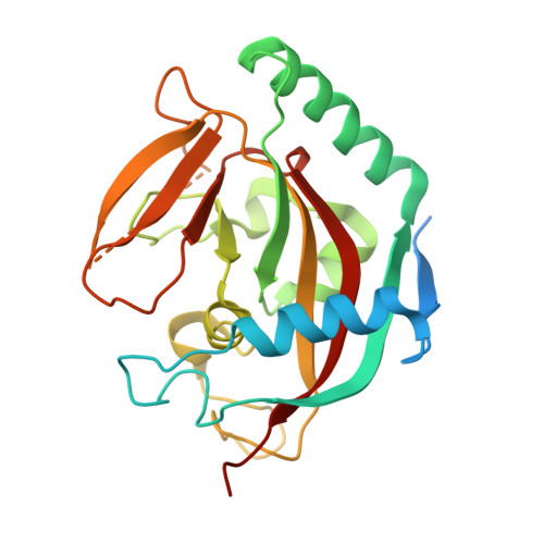
4BUF

Crystal structure of human tankyrase 2 in complex with 2-(4- acetylphenyl)-3,4-dihydroquinazolin-4-one


Experimental Data Snapshot

  • Method: X-RAY DIFFRACTION
  • Resolution: 2.50 Å
  • R-Value Free: 0.262 
  • R-Value Work: 0.200 
  • R-Value Observed: 0.203 

wwPDB Validation   3D Report Full Report


Ligand Structure Quality Assessment 


This is version 1.4 of the entry. See complete history


Literature

Para-Substituted 2-Phenyl-3,4-Dihydroquinazolin-4-Ones as Potent and Selective Tankyrase Inhibitors.

Haikarainen, T.Koivunen, J.Narwal, M.Venkannagari, H.Obaji, E.Joensuu, P.Pihlajaniemi, T.Lehtio, L.

(2013) ChemMedChem 8: 1978

  • DOI: https://doi.org/10.1002/cmdc.201300337
  • Primary Citation of Related Structures:  
    4BU3, 4BU5, 4BU6, 4BU7, 4BU8, 4BU9, 4BUA, 4BUD, 4BUE, 4BUF, 4BUI, 4BUS, 4BUT, 4BUU, 4BUV, 4BUW, 4BUX, 4BUY

  • PubMed Abstract: 

    Human tankyrases are attractive drug targets, especially for the treatment of cancer. We identified a set of highly potent tankyrase inhibitors based on a 2-phenyl-3,4-dihydroquinazolin-4-one scaffold. Substitutions at the para position of the scaffold's phenyl group were evaluated as a strategy to increase potency and improve selectivity. The best compounds displayed single-digit nanomolar potencies, and profiling against several human diphtheria-toxin-like ADP-ribosyltransferases revealed that a subset of these compounds are highly selective tankyrase inhibitors. The compounds also effectively inhibit Wnt signaling in HEK293 cells. The binding mode of all inhibitors was studied by protein X-ray crystallography. This allowed us to establish a structural basis for the development of highly potent and selective tankyrase inhibitors based on the 2-phenyl-3,4-dihydroquinazolin-4-one scaffold and outline a rational approach to the modification of other inhibitor scaffolds that bind to the nicotinamide site of the catalytic domain.


  • Organizational Affiliation

    Department of Biochemistry and Biocenter Oulu, 90014 University of Oulu, PO Box 3000 Oulu (Finland).


Macromolecules
Find similar proteins by:  (by identity cutoff)  |  3D Structure
Entity ID: 1
MoleculeChains Sequence LengthOrganismDetailsImage
TANKYRASE-2
A, B
240Homo sapiensMutation(s): 0 
EC: 2.4.2.30
UniProt & NIH Common Fund Data Resources
Find proteins for Q9H2K2 (Homo sapiens)
Explore Q9H2K2 
Go to UniProtKB:  Q9H2K2
PHAROS:  Q9H2K2
GTEx:  ENSG00000107854 
Entity Groups  
Sequence Clusters30% Identity50% Identity70% Identity90% Identity95% Identity100% Identity
UniProt GroupQ9H2K2
Sequence Annotations
Expand
  • Reference Sequence
Small Molecules
Ligands 4 Unique
IDChains Name / Formula / InChI Key2D Diagram3D Interactions
F36
Query on F36

Download Ideal Coordinates CCD File 
C [auth A],
I [auth B]
2-(4-acetylphenyl)-3,4-dihydroquinazolin-4-one
C16 H12 N2 O2
VLRQJLLWRMXGLP-UHFFFAOYSA-N
SO4
Query on SO4

Download Ideal Coordinates CCD File 
D [auth A],
E [auth A],
J [auth B],
K [auth B]
SULFATE ION
O4 S
QAOWNCQODCNURD-UHFFFAOYSA-L
GOL
Query on GOL

Download Ideal Coordinates CCD File 
G [auth A],
H [auth A]
GLYCEROL
C3 H8 O3
PEDCQBHIVMGVHV-UHFFFAOYSA-N
ZN
Query on ZN

Download Ideal Coordinates CCD File 
F [auth A],
L [auth B]
ZINC ION
Zn
PTFCDOFLOPIGGS-UHFFFAOYSA-N
Binding Affinity Annotations 
IDSourceBinding Affinity
F36 Binding MOAD:  4BUF IC50: 38 (nM) from 1 assay(s)
Experimental Data & Validation

Experimental Data

  • Method: X-RAY DIFFRACTION
  • Resolution: 2.50 Å
  • R-Value Free: 0.262 
  • R-Value Work: 0.200 
  • R-Value Observed: 0.203 
  • Space Group: C 2 2 21
Unit Cell:
Length ( Å )Angle ( ˚ )
a = 91.655α = 90
b = 97.384β = 90
c = 117.545γ = 90
Software Package:
Software NamePurpose
REFMACrefinement
SAINTdata reduction
SADABSdata scaling

Structure Validation

View Full Validation Report


Ligand Structure Quality Assessment 


Entry History 

Deposition Data

Revision History  (Full details and data files)

  • Version 1.0: 2013-10-30
    Type: Initial release
  • Version 1.1: 2013-12-11
    Changes: Database references
  • Version 1.2: 2017-07-05
    Changes: Data collection
  • Version 1.3: 2019-01-30
    Changes: Data collection, Experimental preparation
  • Version 1.4: 2019-02-06
    Changes: Data collection, Experimental preparation