4BHM | pdb_00004bhm

The crystal structure of MoSub1-DNA complex reveals a novel DNA binding mode


Experimental Data Snapshot

  • Method: X-RAY DIFFRACTION
  • Resolution: 2.70 Å
  • R-Value Free: 
    0.243 (Depositor), 0.237 (DCC) 
  • R-Value Work: 
    0.184 (Depositor), 0.192 (DCC) 
  • R-Value Observed: 
    0.187 (Depositor) 

wwPDB Validation 3D Report Full Report

Validation slider image for 4BHM

This is version 1.2 of the entry. See complete history

Literature

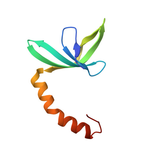
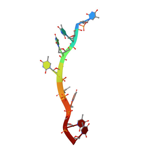
Substitution of Tryptophan 89 with Tyrosine Switches the DNA Binding Mode of Pc4.

Huang, J.Zhao, Y.Liu, H.Huang, D.Cheng, X.Zhao, W.Taylor, I.A.Liu, J.Peng, Y.L.

(2015) Sci Rep 5: 8789

  • DOI: https://doi.org/10.1038/srep08789
  • Primary Citation Related Structures: 
    4BHM, 4USG

  • PubMed Abstract: 

    PC4, a well-known general transcription cofactor, has multiple functions in transcription and DNA repair. Residue W89, is engaged in stacking interactions with DNA in PC4, but substituted by tyrosine in some PC4 orthologous proteins. In order to understand the consequences and reveal the molecular details of this substitution we have determined the crystal structures of the PC4 orthologue MoSub1 and a PC4 W89Y mutant in complex with DNA. In the structure of MoSub1-DNA complex, Y74 interacts directly with a single nucleotide of oligo DNA. By comparison, the equivalent residue, W89 in wild type PC4 interacts with two nucleotides and the base of the second nucleotide has distinct orientation relative to that of the first one. A hydrophobic patch around W89 that favours interaction with two nucleotides is not formed in the PC4 W89Y mutant. Therefore, the change of the surface hydrophobicity around residue 89 results in a difference between the modes of DNA interaction. These results indicate that the conserved Y74 in MoSub1 or W89 in PC4, are not only key residues in making specific interactions with DNA but also required to determine the DNA binding mode of PC4 proteins.


  • Organizational Affiliation
    • 1] State key Laboratory of Agrobiotechnology, China Agricultural University, No2 Yunamingyuanxilu, Beijing. 100193, China [2] MOA Key Laboratory of Plant Pathology, China Agricultural University, No2 Yunamingyuanxilu, Beijing. 100193, China [3] College of Agronomy and Plant Protection, Qingdao Agricultural University, Qingdao, Shandong. 266109, China.

Macromolecule Content 

  • Total Structure Weight: 60.9 kDa 
  • Atom Count: 4,141 
  • Modeled Residue Count: 486 
  • Deposited Residue Count: 498 
  • Unique protein chains: 1
  • Unique nucleic acid chains: 1

Macromolecules


Find similar proteins by:|  3D Structure
Entity ID: 1
MoleculeChains  Sequence LengthOrganismDetailsImage
MOSUB1 TRANSCRIPTION COFACTOR
A, B, C, D, E
A, B, C, D, E, F
79Pyricularia oryzaeMutation(s): 0 
UniProt
Find proteins for G4NEJ8 (Pyricularia oryzae (strain 70-15 / ATCC MYA-4617 / FGSC 8958))
Explore G4NEJ8 
Go to UniProtKB:  G4NEJ8
Entity Groups
Sequence Clusters30% Identity50% Identity70% Identity90% Identity95% Identity100% Identity
UniProt GroupG4NEJ8
Sequence Annotations
Expand
Reference Sequence
Find similar nucleic acids by:  Sequence
Entity ID: 2
MoleculeChains LengthOrganismImage
5'-D(*TP*TP*TP*TP*TP*TP*TP*TP)-3'
G, H, I
8Pyricularia oryzae
Sequence Annotations
Expand
Reference Sequence

Experimental Data & Validation

Experimental Data

  • Method: X-RAY DIFFRACTION
  • Resolution: 2.70 Å
  • R-Value Free:  0.243 (Depositor), 0.237 (DCC) 
  • R-Value Work:  0.184 (Depositor), 0.192 (DCC) 
  • R-Value Observed: 0.187 (Depositor) 
Space Group: P 1 21 1
Unit Cell:
Length ( Å )Angle ( ˚ )
a = 84.19α = 90
b = 57.97β = 107.02
c = 83.76γ = 90
Software Package:
Software NamePurpose
PHENIXrefinement

Structure Validation

View Full Validation Report


Entry History 

Deposition Data

Revision History  (Full details and data files)

  • Version 1.0: 2014-04-23
    Type: Initial release
  • Version 1.1: 2015-03-18
    Changes: Database references
  • Version 1.2: 2024-05-08
    Changes: Data collection, Database references, Derived calculations, Other