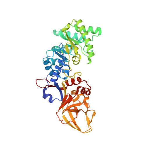
4BEU

Structure of Vibrio cholerae broad spectrum racemase


Experimental Data Snapshot

  • Method: X-RAY DIFFRACTION
  • Resolution: 1.15 Å
  • R-Value Free: 0.173 
  • R-Value Work: 0.163 
  • R-Value Observed: 0.164 

Starting Model: experimental
View more details

wwPDB Validation   3D Report Full Report


Ligand Structure Quality Assessment 


This is version 2.1 of the entry. See complete history


Literature

Structural Basis for the Broad Specificity of a New Family of Amino-Acid Racemases.

Espaillat, A.Carrasco-Lopez, C.Bernardo-Garcia, N.Pietrosemoli, N.Otero, L.H.Alvarez, L.De Pedro, M.A.Pazos, F.Davis, B.M.Waldor, M.K.Hermoso, J.A.Cava, F.

(2014) Acta Crystallogr D Biol Crystallogr 70: 79

  • DOI: https://doi.org/10.1107/S1399004713024838
  • Primary Citation of Related Structures:  
    4BEQ, 4BEU, 4BF5, 4BHY

  • PubMed Abstract: 

    Broad-spectrum amino-acid racemases (Bsrs) enable bacteria to generate noncanonical D-amino acids, the roles of which in microbial physiology, including the modulation of cell-wall structure and the dissolution of biofilms, are just beginning to be appreciated. Here, extensive crystallographic, mutational, biochemical and bioinformatic studies were used to define the molecular features of the racemase BsrV that enable this enzyme to accommodate more diverse substrates than the related PLP-dependent alanine racemases. Conserved residues were identified that distinguish BsrV and a newly defined family of broad-spectrum racemases from alanine racemases, and these residues were found to be key mediators of the multispecificity of BrsV. Finally, the structural analysis of an additional Bsr that was identified in the bioinformatic analysis confirmed that the distinguishing features of BrsV are conserved among Bsr family members.


  • Organizational Affiliation

    Centro de Biología Molecular `Severo Ochoa', Universidad Autónoma de Madrid-Consejo Superior de Investigaciones Científicas (CSIC), 28049 Madrid, Spain.


Macromolecules
Find similar proteins by:  (by identity cutoff)  |  3D Structure
Entity ID: 1
MoleculeChains Sequence LengthOrganismDetailsImage
ALANINE RACEMASE392Vibrio choleraeMutation(s): 0 
EC: 5.1.1.1 (PDB Primary Data), 5.1.1.10 (UniProt)
UniProt
Find proteins for Q9KSE5 (Vibrio cholerae serotype O1 (strain ATCC 39315 / El Tor Inaba N16961))
Explore Q9KSE5 
Go to UniProtKB:  Q9KSE5
Entity Groups  
Sequence Clusters30% Identity50% Identity70% Identity90% Identity95% Identity100% Identity
UniProt GroupQ9KSE5
Sequence Annotations
Expand
  • Reference Sequence
Experimental Data & Validation

Experimental Data

  • Method: X-RAY DIFFRACTION
  • Resolution: 1.15 Å
  • R-Value Free: 0.173 
  • R-Value Work: 0.163 
  • R-Value Observed: 0.164 
  • Space Group: C 1 2 1
Unit Cell:
Length ( Å )Angle ( ˚ )
a = 96.621α = 90
b = 51.093β = 101.15
c = 76.731γ = 90
Software Package:
Software NamePurpose
PHENIXrefinement
XDSdata reduction
SCALAdata scaling
MOLREPphasing

Structure Validation

View Full Validation Report


Ligand Structure Quality Assessment 


Entry History 

Deposition Data

Revision History  (Full details and data files)

  • Version 1.0: 2014-01-15
    Type: Initial release
  • Version 1.1: 2014-01-22
    Changes: Database references
  • Version 1.2: 2014-07-09
    Changes: Database references
  • Version 2.0: 2020-01-15
    Changes: Atomic model, Derived calculations, Other
  • Version 2.1: 2023-12-20
    Changes: Data collection, Database references, Derived calculations, Refinement description