4BC6 | pdb_00004bc6

Crystal structure of human serine threonine kinase-10 bound to novel Bosutinib Isoform 1, previously thought to be Bosutinib


Experimental Data Snapshot

  • Method: X-RAY DIFFRACTION
  • Resolution: 2.20 Å
  • R-Value Free: 
    0.254 (Depositor), 0.281 (DCC) 
  • R-Value Work: 
    0.211 (Depositor), 0.233 (DCC) 
  • R-Value Observed: 
    0.214 (Depositor) 

Starting Model: experimental
View more details

wwPDB Validation 3D Report Full Report

Validation slider image for 4BC6

Ligand Structure Quality Assessment 


This is version 1.2 of the entry. See complete history

Literature

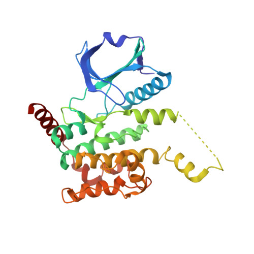
Crystal Structure of Human Serine Threonine Kinase- 10 Bound to Novel Bosutinib Isoform 1, Previously Thought to be Bosutinib

Vollmar, M.Szklarz, M.Chaikuad, A.Elkins, J.Savitsky, P.Azeez, K.A.Salah, E.Krojer, T.Canning, P.Muniz, J.R.C.Bountra, C.Arrowsmith, C.H.Weigelt, J.Edwards, A.von Delft, F.Knapp, S.

To be published.

Macromolecule Content 

  • Total Structure Weight: 33.72 kDa 
  • Atom Count: 2,190 
  • Modeled Residue Count: 285 
  • Deposited Residue Count: 293 
  • Unique protein chains: 1

Macromolecules

Find similar proteins by:|  3D Structure
Entity ID: 1
MoleculeChains  Sequence LengthOrganismDetailsImage
SERINE/THREONINE-PROTEIN KINASE 10293Homo sapiensMutation(s): 0 
EC: 2.7.11.1
UniProt & NIH Common Fund Data Resources
Find proteins for O94804 (Homo sapiens)
Explore O94804 
Go to UniProtKB:  O94804
PHAROS:  O94804
GTEx:  ENSG00000072786 
Entity Groups
Sequence Clusters30% Identity50% Identity70% Identity90% Identity95% Identity100% Identity
UniProt GroupO94804
Sequence Annotations
Expand
Reference Sequence

Small Molecules

Ligands 2 Unique
IDChains Name / Formula / InChI Key2D Diagram3D Interactions
XZN

Query on XZN


Download:Ideal Coordinates CCD File
B [auth A]4-[(3,5-DICHLORO-4-METHOXYPHENYL)AMINO]-6-METHOXY-7-[3-(4-METHYLPIPERAZIN-1-YL)PROPOXY]QUINOLINE-3-CARBONITRILE
C26 H29 Cl2 N5 O3
YCLIWTLPTXAGPQ-UHFFFAOYSA-N
EDO

Query on EDO


Download:Ideal Coordinates CCD File
C [auth A]1,2-ETHANEDIOL
C2 H6 O2
LYCAIKOWRPUZTN-UHFFFAOYSA-N

Experimental Data & Validation

Experimental Data

  • Method: X-RAY DIFFRACTION
  • Resolution: 2.20 Å
  • R-Value Free:  0.254 (Depositor), 0.281 (DCC) 
  • R-Value Work:  0.211 (Depositor), 0.233 (DCC) 
  • R-Value Observed: 0.214 (Depositor) 
Space Group: I 2 2 2
Unit Cell:
Length ( Å )Angle ( ˚ )
a = 51.17α = 90
b = 114.03β = 90
c = 134.34γ = 90
Software Package:
Software NamePurpose
BUSTERrefinement
XDSdata reduction
SCALAdata scaling
PHASERphasing

Structure Validation

View Full Validation Report


Ligand Structure Quality Assessment 


Entry History 

Revision History  (Full details and data files)

  • Version 1.0: 2012-10-10
    Type: Initial release
  • Version 1.1: 2018-01-24
    Changes: Database references, Structure summary
  • Version 1.2: 2023-12-20
    Changes: Data collection, Database references, Derived calculations, Other, Refinement description