Watching a Signaling Protein Function in Real Time Via 100-Ps Time-Resolved Laue Crystallography.
Schotte, F., Cho, H.S., Kaila, V.R.I., Kamikubo, H., Dashdorj, N., Henry, E.R., Graber, T.J., Henning, R., Wulff, M., Hummer, G., Kataoka, M., Anfinrud, P.A.(2012) Proc Natl Acad Sci U S A 109: 19256
- PubMed: 23132943 Search on PubMedSearch on PubMed Central
- DOI: https://doi.org/10.1073/pnas.1210938109
- Primary Citation Related Structures:
4B9O, 4BBT, 4BBU, 4BBV - PubMed Abstract:
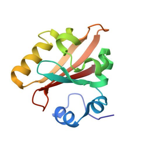
To understand how signaling proteins function, it is crucial to know the time-ordered sequence of events that lead to the signaling state. We recently developed on the BioCARS 14-IDB beamline at the Advanced Photon Source the infrastructure required to characterize structural changes in protein crystals with near-atomic spatial resolution and 150-ps time resolution, and have used this capability to track the reversible photocycle of photoactive yellow protein (PYP) following trans-to-cis photoisomerization of its p-coumaric acid (pCA) chromophore over 10 decades of time. The first of four major intermediates characterized in this study is highly contorted, with the pCA carbonyl rotated nearly 90° out of the plane of the phenolate. A hydrogen bond between the pCA carbonyl and the Cys69 backbone constrains the chromophore in this unusual twisted conformation. Density functional theory calculations confirm that this structure is chemically plausible and corresponds to a strained cis intermediate. This unique structure is short-lived (∼600 ps), has not been observed in prior cryocrystallography experiments, and is the progenitor of intermediates characterized in previous nanosecond time-resolved Laue crystallography studies. The structural transitions unveiled during the PYP photocycle include trans/cis isomerization, the breaking and making of hydrogen bonds, formation/relaxation of strain, and gated water penetration into the interior of the protein. This mechanistically detailed, near-atomic resolution description of the complete PYP photocycle provides a framework for understanding signal transduction in proteins, and for assessing and validating theoretical/computational approaches in protein biophysics.
- Laboratory of Chemical Physics, National Institute of Diabetes and Digestive and Kidney Diseases, National Institutes of Health, Bethesda, MD 20892, USA.
Organizational Affiliation: