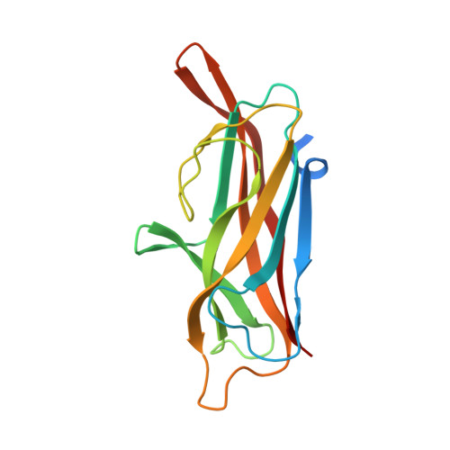
4B9G

Structure of CssB subunit complemented with donor strand from CssA subunit of enterotoxigenic Escherichia coli colonization factor CS6


Experimental Data Snapshot

  • Method: X-RAY DIFFRACTION
  • Resolution: 1.04 Å
  • R-Value Free: 0.167 
  • R-Value Work: 0.155 
  • R-Value Observed: 0.156 

wwPDB Validation   3D Report Full Report


This is version 1.3 of the entry. See complete history


Literature

Crystal Structure of Enterotoxigenic Escherichia Coli Colonization Factor Cs6 Reveals a Novel Type of Functional Assembly.

Roy, S.P.Rahman, M.M.Yu, X.D.Tuittila, M.Knight, S.D.Zavialov, A.V.

(2012) Mol Microbiol 86: 1100

  • DOI: https://doi.org/10.1111/mmi.12044
  • Primary Citation of Related Structures:  
    4B9G, 4B9I, 4B9J

  • PubMed Abstract: 

    Coli surface antigen 6 (CS6) is a widely expressed enterotoxigenic Escherichia coli (ETEC) colonization factor that mediates bacterial attachment to the small intestinal epithelium. CS6 is a polymer of two protein subunits CssA and CssB, which are secreted and assembled on the cell surface via the CssC/CssD chaperone usher (CU) pathway. Here, we present an atomic resolution model for the structure of CS6 based on the results of X-ray crystallographic, spectroscopic and biochemical studies, and suggest a mechanism for CS6-mediated adhesion. We show that the CssA and CssB subunits are assembled alternately in linear fibres by the principle of donor strand complementation. This type of fibre assembly is novel for CU assembled adhesins. We also show that both subunits in the fibre bind to receptors on epithelial cells, and that CssB, but not CssA, specifically recognizes the extracellular matrix protein fibronectin. Taken together, structural and functional results suggest that CS6 is an adhesive organelle of a novel type, a hetero-polyadhesin that is capable of polyvalent attachment to different receptors.


  • Organizational Affiliation

    Department of Molecular Biology, Uppsala BioCenter, Swedish University of Agricultural Sciences, SE-753 24 Uppsala, Sweden.


Macromolecules
Find similar proteins by:  (by identity cutoff)  |  3D Structure
Entity ID: 1
MoleculeChains Sequence LengthOrganismDetailsImage
CS6 FIMBRIAL SUBUNIT B, CS6 FIMBRIAL SUBUNIT A
A, B
161Escherichia coliMutation(s): 0 
UniProt
Find proteins for P53510 (Escherichia coli)
Explore P53510 
Go to UniProtKB:  P53510
Entity Groups  
Sequence Clusters30% Identity50% Identity70% Identity90% Identity95% Identity100% Identity
UniProt GroupP53510
Sequence Annotations
Expand
  • Reference Sequence
Experimental Data & Validation

Experimental Data

  • Method: X-RAY DIFFRACTION
  • Resolution: 1.04 Å
  • R-Value Free: 0.167 
  • R-Value Work: 0.155 
  • R-Value Observed: 0.156 
  • Space Group: P 21 21 21
Unit Cell:
Length ( Å )Angle ( ˚ )
a = 51.956α = 90
b = 69.225β = 90
c = 72.359γ = 90
Software Package:
Software NamePurpose
PHENIXrefinement
MOSFLMdata reduction
SCALAdata scaling
PHENIXphasing

Structure Validation

View Full Validation Report


Entry History 

Deposition Data

Revision History  (Full details and data files)

  • Version 1.0: 2012-11-07
    Type: Initial release
  • Version 1.1: 2012-12-05
    Changes: Atomic model
  • Version 1.2: 2012-12-12
    Changes: Database references
  • Version 1.3: 2024-05-08
    Changes: Data collection, Database references, Other