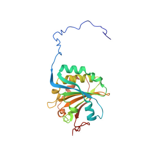
4B6U

Solution structure of eIF4E3 in complex with m7GDP


Experimental Data Snapshot

  • Method: SOLUTION NMR
  • Conformers Calculated: 100 
  • Conformers Submitted: 10 
  • Selection Criteria: LEAST RESTRAINT VIOLATION 

wwPDB Validation   3D Report Full Report


This is version 2.0 of the entry. See complete history


Literature

Eif4E3 Acts as a Tumor Suppressor by Utilizing an Atypical Mode of Methyl-7-Guanosine CAP Recognition

Osborne, M.J.Volpon, L.Kornblatt, J.A.Culjkovic-Kraljcic, B.Baguet, A.Borden, K.L.B.

(2013) Proc Natl Acad Sci U S A 110: 3877

  • DOI: https://doi.org/10.1073/pnas.1216862110
  • Primary Citation of Related Structures:  
    4B6U, 4B6V

  • PubMed Abstract: 

    Recognition of the methyl-7-guanosine (m(7)G) cap structure on mRNA is an essential feature of mRNA metabolism and thus gene expression. Eukaryotic translation initiation factor 4E (eIF4E) promotes translation, mRNA export, proliferation, and oncogenic transformation dependent on this cap-binding activity. eIF4E-cap recognition is mediated via complementary charge interactions of the positively charged m(7)G cap between the negative π-electron clouds from two aromatic residues. Here, we demonstrate that a variant subfamily, eIF4E3, specifically binds the m(7)G cap in the absence of an aromatic sandwich, using instead a different spatial arrangement of residues to provide the necessary electrostatic and van der Waals contacts. Contacts are much more extensive between eIF4E3-cap than other family members. Structural analyses of other cap-binding proteins indicate this recognition mode is atypical. We demonstrate that eIF4E3 relies on this cap-binding activity to act as a tumor suppressor, competing with the growth-promoting functions of eIF4E. In fact, reduced eIF4E3 in high eIF4E cancers suggests that eIF4E3 underlies a clinically relevant inhibitory mechanism that is lost in some malignancies. Taken together, there is more structural plasticity in cap recognition than previously thought, and this is physiologically relevant.


  • Organizational Affiliation

    Institute for Research in Immunology and Cancer, Department of Pathology and Cell Biology, Université de Montréal, Montreal, QC, Canada H3T 1J4.


Macromolecules
Find similar proteins by:  (by identity cutoff)  |  3D Structure
Entity ID: 1
MoleculeChains Sequence LengthOrganismDetailsImage
EUKARYOTIC TRANSLATION INITIATION FACTOR 4E TYPE 3207Mus musculusMutation(s): 0 
UniProt & NIH Common Fund Data Resources
Find proteins for Q9DBB5 (Mus musculus)
Explore Q9DBB5 
Go to UniProtKB:  Q9DBB5
IMPC:  MGI:1914142
Entity Groups  
Sequence Clusters30% Identity50% Identity70% Identity90% Identity95% Identity100% Identity
UniProt GroupQ9DBB5
Sequence Annotations
Expand
  • Reference Sequence
Small Molecules
Ligands 1 Unique
IDChains Name / Formula / InChI Key2D Diagram3D Interactions
M7G
Query on M7G

Download Ideal Coordinates CCD File 
B [auth A]7N-METHYL-8-HYDROGUANOSINE-5'-DIPHOSPHATE
C11 H18 N5 O11 P2
SBASPRRECYVBRF-KQYNXXCUSA-O
Binding Affinity Annotations 
IDSourceBinding Affinity
M7G PDBBind:  4B6U Kd: 7700 (nM) from 1 assay(s)
Experimental Data & Validation

Experimental Data

  • Method: SOLUTION NMR
  • Conformers Calculated: 100 
  • Conformers Submitted: 10 
  • Selection Criteria: LEAST RESTRAINT VIOLATION 

Structure Validation

View Full Validation Report


Entry History 

Deposition Data

Revision History  (Full details and data files)

  • Version 1.0: 2013-03-06
    Type: Initial release
  • Version 1.1: 2013-03-20
    Changes: Database references
  • Version 1.2: 2016-05-04
    Changes: Atomic model, Other
  • Version 1.3: 2018-01-17
    Changes: Structure summary
  • Version 1.4: 2023-06-14
    Changes: Data collection, Database references, Derived calculations, Other
  • Version 2.0: 2024-06-19
    Changes: Data collection, Database references, Non-polymer description, Structure summary