4B3H | pdb_00004b3h

Crystal structure of Mycobacterium tuberculosis fatty acid beta- oxidation complex


Experimental Data Snapshot

  • Method: X-RAY DIFFRACTION
  • Resolution: 2.30 Å
  • R-Value Free: 
    0.224 (Depositor), 0.224 (DCC) 
  • R-Value Work: 
    0.195 (Depositor), 0.195 (DCC) 
  • R-Value Observed: 
    0.197 (Depositor) 

Starting Model: experimental
View more details

wwPDB Validation 3D Report Full Report

Validation slider image for 4B3H

This is version 1.2 of the entry. See complete history

Literature

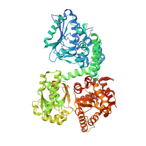
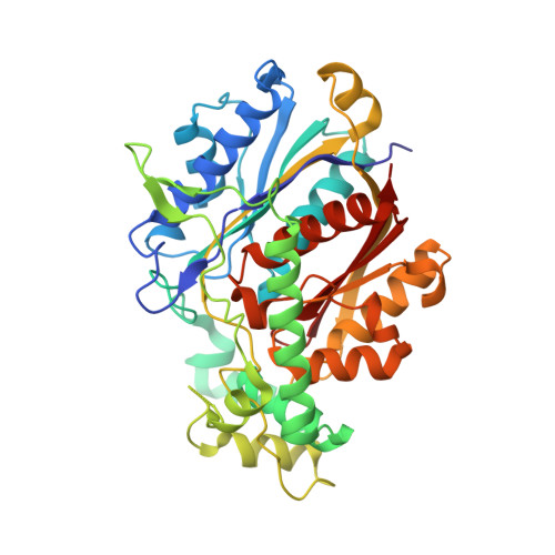
Structure of Mycobacterial Beta-Oxidation Trifunctional Enzyme Reveals its Altered Assembly and Putative Substrate Channeling Pathway.

Venkatesan, R.Wierenga, R.K.

(2013) ACS Chem Biol 8: 1063

  • DOI: https://doi.org/10.1021/cb400007k
  • Primary Citation Related Structures: 
    4B3H, 4B3I, 4B3J

  • PubMed Abstract: 

    The incidence of tuberculosis is increasing due to the appearance of new drug-resistant variants. A thorough understanding of the disease organism is essential in order to create more effective drugs. In an attempt to understand better the poorly studied lipid metabolism of Mycobacterium tuberculosis (Mtb), we identified and characterized its fatty acid β-oxidation complex (trifunctional enzyme (TFE)). TFE is an α(2)β(2) complex consisting of two types of polypeptides catalyzing three of the four reactions of the β-oxidation of fatty acids. The kinetic constants (k(cat) and K(m)) show that the complexed α chain is more active than the individual α chain. Crystal structures of Mtb TFE (mtTFE) reveal that the quaternary assembly is strikingly different from the already known Pseudomonas fragi TFE (pfTFE) assembly due to the presence of a helical insertion (LA5) in the mtTFE-β subunit. This helical insertion prevents the pfTFE mode of assembly, as it would clash with helix H9A of the TFE-α chain. The mtTFE assembly appears to be more rigid and results in a different substrate channeling path between the α and the β subunits. Structural comparisons suggest that the mtTFE active sites can accommodate bulkier fatty acyl chains than in pfTFE. Although another thiolase (FadA2), more closely related to human TFE-β/thiolase, is present in the Mtb genome, it does not form a complex with mtTFE-α. Extensive phylogenetic analyses show that there are at least four TFE subfamilies. Our studies highlight the molecular properties of mtTFE, significantly extending the structural knowledge on this type of very interesting multifunctional enzymes.


  • Organizational Affiliation
    • Department of Biochemistry and Biocenter Oulu, University of Oulu, Oulu 90014, Finland.

Macromolecule Content 

  • Total Structure Weight: 244.64 kDa 
  • Atom Count: 18,048 
  • Modeled Residue Count: 2,256 
  • Deposited Residue Count: 2,278 
  • Unique protein chains: 2

Macromolecules

Find similar proteins by:|  3D Structure
Entity ID: 1
MoleculeChains  Sequence LengthOrganismDetailsImage
FATTY ACID BETA-OXIDATION COMPLEX ALPHA-CHAIN FADB
A, B
736Mycobacterium tuberculosis H37RvMutation(s): 0 
EC: 4.2.1.17 (PDB Primary Data), 1.1.1.35 (PDB Primary Data)
UniProt
Find proteins for O53872 (Mycobacterium tuberculosis (strain ATCC 25618 / H37Rv))
Explore O53872 
Go to UniProtKB:  O53872
Entity Groups
Sequence Clusters30% Identity50% Identity70% Identity90% Identity95% Identity100% Identity
UniProt GroupO53872
Sequence Annotations
Expand
Reference Sequence
Find similar proteins by:|  3D Structure
Entity ID: 2
MoleculeChains  Sequence LengthOrganismDetailsImage
FATTY ACID BETA-OXIDATION COMPLEX BETA-CHAIN FADA
C, D
403Mycobacterium tuberculosis H37RvMutation(s): 0 
EC: 2.3.1.9 (PDB Primary Data), 2.3.1 (UniProt)
UniProt
Find proteins for O53871 (Mycobacterium tuberculosis (strain ATCC 25618 / H37Rv))
Explore O53871 
Go to UniProtKB:  O53871
Entity Groups
Sequence Clusters30% Identity50% Identity70% Identity90% Identity95% Identity100% Identity
UniProt GroupO53871
Sequence Annotations
Expand
Reference Sequence

Small Molecules

Ligands 2 Unique
IDChains Name / Formula / InChI Key2D Diagram3D Interactions
SO4

Query on SO4


Download:Ideal Coordinates CCD File
BA [auth C]
CA [auth C]
DA [auth C]
E [auth A]
EA [auth C]
BA [auth C],
CA [auth C],
DA [auth C],
E [auth A],
EA [auth C],
F [auth A],
FA [auth C],
G [auth A],
GA [auth C],
H [auth A],
HA [auth C],
I [auth A],
IA [auth C],
J [auth A],
K [auth A],
KA [auth D],
LA [auth D],
MA [auth D],
NA [auth D],
OA [auth D],
P [auth B],
PA [auth D],
Q [auth B],
R [auth B],
S [auth B],
T [auth B],
U [auth B],
V [auth B],
W [auth B],
X [auth B]
SULFATE ION
O4 S
QAOWNCQODCNURD-UHFFFAOYSA-L
GOL

Query on GOL


Download:Ideal Coordinates CCD File
AA [auth B]
JA [auth C]
L [auth A]
M [auth A]
N [auth A]
AA [auth B],
JA [auth C],
L [auth A],
M [auth A],
N [auth A],
O [auth A],
QA [auth D],
Y [auth B],
Z [auth B]
GLYCEROL
C3 H8 O3
PEDCQBHIVMGVHV-UHFFFAOYSA-N

Experimental Data & Validation

Experimental Data

  • Method: X-RAY DIFFRACTION
  • Resolution: 2.30 Å
  • R-Value Free:  0.224 (Depositor), 0.224 (DCC) 
  • R-Value Work:  0.195 (Depositor), 0.195 (DCC) 
  • R-Value Observed: 0.197 (Depositor) 
Space Group: C 1 2 1
Unit Cell:
Length ( Å )Angle ( ˚ )
a = 249.46α = 90
b = 134.25β = 110.74
c = 118.4γ = 90
Software Package:
Software NamePurpose
REFMACrefinement
MOSFLMdata reduction
SCALAdata scaling
PHASERphasing

Structure Validation

View Full Validation Report


Entry History 

Deposition Data

Revision History  (Full details and data files)

  • Version 1.0: 2013-03-27
    Type: Initial release
  • Version 1.1: 2013-05-29
    Changes: Database references
  • Version 1.2: 2023-12-20
    Changes: Data collection, Database references, Derived calculations, Other, Refinement description