4AY6 | pdb_00004ay6

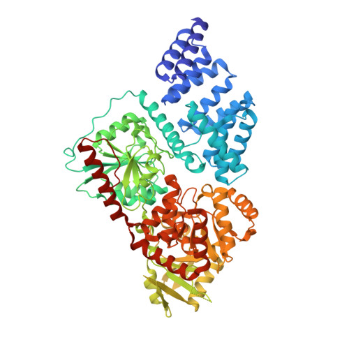
Human O-GlcNAc transferase (OGT) in complex with UDP-5SGlcNAc and substrate peptide


Experimental Data Snapshot

  • Method: X-RAY DIFFRACTION
  • Resolution: 3.30 Å
  • R-Value Free: 
    0.272 (Depositor), 0.272 (DCC) 
  • R-Value Work: 
    0.227 (Depositor), 0.226 (DCC) 
  • R-Value Observed: 
    0.227 (Depositor) 

wwPDB Validation 3D Report Full Report

Validation slider image for 4AY6

Ligand Structure Quality Assessment 


This is version 1.3 of the entry. See complete history. 

Literature

O-Glcnac Transferase Invokes Nucleotide Sugar Pyrophosphate Participation in Catalysis

Schimpl, M., Zheng, X., Borodkin, V.S., Blair, D.E., Ferenbach, A.T., Schuettelkopf, A.W., Navratilova, I., Aristotelous, T., Albarbarawi, O., Robinson, D.A., Macnaughtan, M.A., Van Aalten, D.M.F.

(2012) Nat Chem Biol 8: 969

  • DOI: https://doi.org/10.1038/nchembio.1108
  • Primary Citation Related Structures: 
    4AY5, 4AY6

  • PubMed Abstract: 

    Protein O-GlcNAcylation is an essential post-translational modification on hundreds of intracellular proteins in metazoa, catalyzed by O-linked β-N-acetylglucosamine (O-GlcNAc) transferase (OGT) using unknown mechanisms of transfer and substrate recognition. Through crystallographic snapshots and mechanism-inspired chemical probes, we define how human OGT recognizes the sugar donor and acceptor peptide and uses a new catalytic mechanism of glycosyl transfer, involving the sugar donor α-phosphate as the catalytic base as well as an essential lysine. This mechanism seems to be a unique evolutionary solution to the spatial constraints imposed by a bulky protein acceptor substrate and explains the unexpected specificity of a recently reported metabolic OGT inhibitor.


  • Organizational Affiliation: 
    • College of Life Sciences, University of Dundee, Dundee, UK.

Macromolecule Content 

  • Total Structure Weight: 332.01 kDa 
  • Atom Count: 22,544 
  • Modeled Residue Count: 2,836 
  • Deposited Residue Count: 2,944 
  • Unique protein chains: 2

Macromolecules

Find similar proteins by:|  3D Structure
Entity ID: 1
MoleculeChains  Sequence LengthOrganismDetailsImage
UDP-N-ACETYLGLUCOSAMINE--PEPTIDE N-ACETYLGLUCOSAMINYLTRANS FERASE 110 KDA SUBUNIT
A, B, C, D
723Homo sapiensMutation(s): 0 
EC: 2.4.1.255
UniProt & NIH Common Fund Data Resources
Find proteins for O15294 (Homo sapiens)
Explore O15294 
Go to UniProtKB:  O15294
PHAROS:  O15294
GTEx:  ENSG00000147162 
Entity Groups
Sequence Clusters30% Identity50% Identity70% Identity90% Identity95% Identity100% Identity
UniProt GroupO15294
Sequence Annotations
Expand
Reference Sequence
Find similar proteins by:  Sequence   |   3D Structure  
Entity ID: 2
MoleculeChains  Sequence LengthOrganismDetailsImage
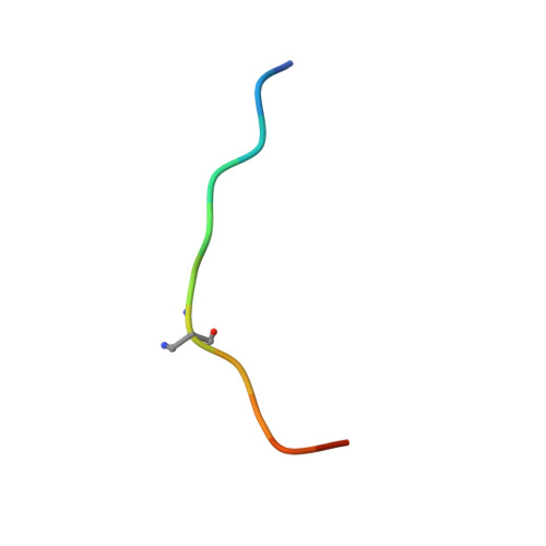
TGF-BETA-ACTIVATED KINASE 1 AND MAP3K7-BINDING PROTEIN 1
E, F, G, H
13Homo sapiensMutation(s): 0 
UniProt & NIH Common Fund Data Resources
Find proteins for Q15750 (Homo sapiens)
Explore Q15750 
Go to UniProtKB:  Q15750
PHAROS:  Q15750
GTEx:  ENSG00000100324 
Entity Groups
Sequence Clusters30% Identity50% Identity70% Identity90% Identity95% Identity100% Identity
UniProt GroupQ15750
Sequence Annotations
Expand
Reference Sequence

Small Molecules

Ligands 2 Unique
IDChains Name / Formula / InChI Key2D Diagram3D Interactions
12V

Query on 12V


Download:Ideal Coordinates CCD File
J [auth A],
L [auth B],
N [auth C],
P [auth D]
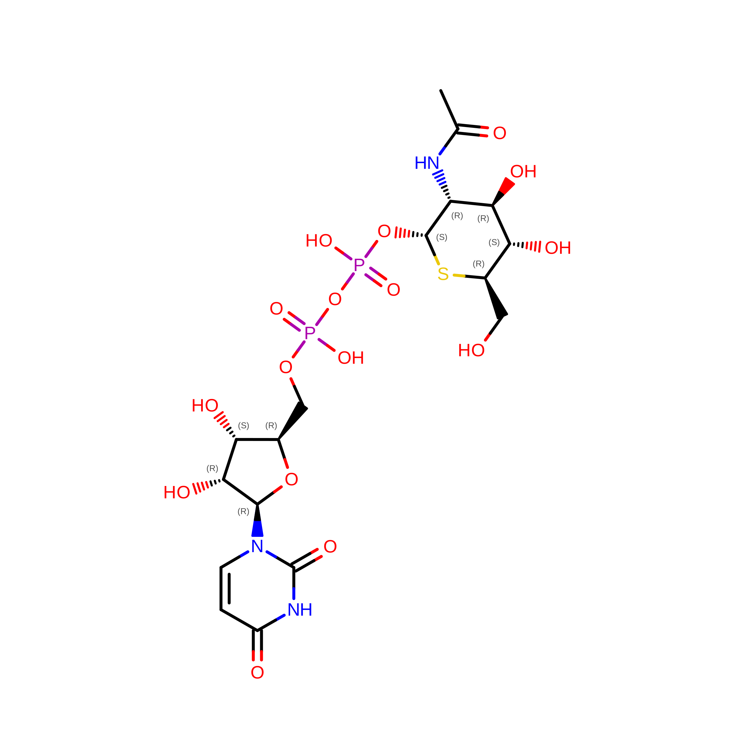
(2S,3R,4R,5S,6R)-3-(acetylamino)-4,5-dihydroxy-6-(hydroxymethyl)tetrahydro-2H-thiopyran-2-yl [(2R,3S,4R,5R)-5-(2,4-dioxo-3,4-dihydropyrimidin-1(2H)-yl)-3,4-dihydroxytetrahydrofuran-2-yl]methyl dihydrogen diphosphate
C17 H27 N3 O16 P2 S
JPRVHSQHWXZSNC-UBDZBXRQSA-N
SO4

Query on SO4


Download:Ideal Coordinates CCD File
I [auth A],
K [auth B],
M [auth C],
O [auth D]
SULFATE ION
O4 S
QAOWNCQODCNURD-UHFFFAOYSA-L
Modified Residues  1 Unique
IDChains TypeFormula2D DiagramParent
DNP
Query on DNP
E, F, G, H
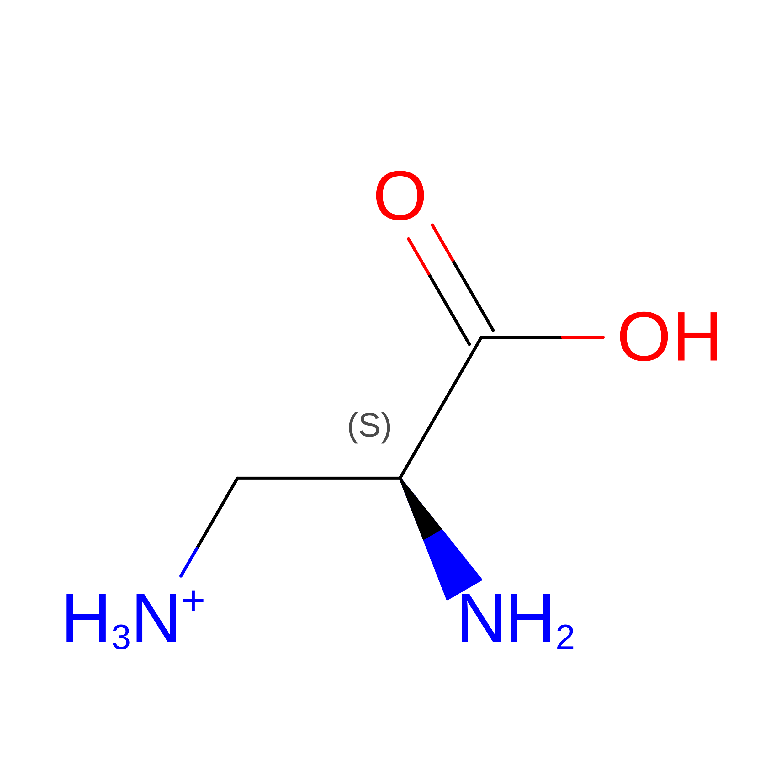
L-PEPTIDE LINKINGC3 H9 N2 O2ALA

Experimental Data & Validation

Experimental Data

  • Method: X-RAY DIFFRACTION
  • Resolution: 3.30 Å
  • R-Value Free:  0.272 (Depositor), 0.272 (DCC) 
  • R-Value Work:  0.227 (Depositor), 0.226 (DCC) 
  • R-Value Observed: 0.227 (Depositor) 
Space Group: P 3 2 1
Unit Cell:
Length ( Å )Angle ( ˚ )
a = 275.439α = 90
b = 275.439β = 90
c = 142.632γ = 120
Software Package:
Software NamePurpose
REFMACrefinement

Structure Validation

View Full Validation Report


Ligand Structure Quality Assessment 


Entry History 

Revision History  (Full details and data files)

  • Version 1.0: 2012-10-24
    Type: Initial release
  • Version 1.1: 2012-12-05
    Changes: Database references
  • Version 1.2: 2012-12-12
    Changes: Database references
  • Version 1.3: 2025-04-09
    Changes: Data collection, Database references, Derived calculations, Other, Structure summary