4AXQ | pdb_00004axq

CRYSTAL STRUCTURE OF ARCHAEMETZINCIN (AMZA) FROM ARCHAEOGLOBUS FULGIDUS AT 1.4 A RESOLUTION


Experimental Data Snapshot

  • Method: X-RAY DIFFRACTION
  • Resolution: 1.40 Å
  • R-Value Free: 
    0.186 (Depositor), 0.192 (DCC) 
  • R-Value Work: 
    0.164 (Depositor), 0.170 (DCC) 
  • R-Value Observed: 
    0.165 (Depositor) 

Starting Model: experimental
View more details

wwPDB Validation 3D Report Full Report

Validation slider image for 4AXQ

This is version 1.3 of the entry. See complete history

Literature

Crystal Structures of Archaemetzincin Reveal a Moldable Substrate-Binding Site.

Graef, C.Schacherl, M.Waltersperger, S.Baumann, U.

(2012) PLoS One 7: 43863

  • DOI: https://doi.org/10.1371/journal.pone.0043863
  • Primary Citation Related Structures: 
    3ZVS, 4A3W, 4AXQ

  • PubMed Abstract: 

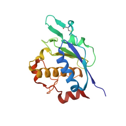
    Archaemetzincins are metalloproteases occurring in archaea and some mammalia. They are distinct from all the other metzincins by their extended active site consensus sequence HEXXHXXGXXHCX(4)CXMX(17)CXXC featuring four conserved cysteine residues. Very little is known about their biological importance and structure-function relationships. Here we present three crystal structures of the archaemetzincin AfAmzA (Uniprot O29917) from Archaeoglobus fulgidus, revealing a metzincin architecture featuring a zinc finger-like structural element involving the conserved cysteines of the consensus motif. The active sites in all three structures are occluded to different extents rendering the enzymes proteolytically inactive against a large variety of tested substrates. Owing to the different ligand binding there are significant differences in active site architecture, revealing a large flexibility of the loops covering the active site cleft. The crystal structures of AfAmzA provide the structural basis for the lack of activity in standard proteolytic assays and imply a triggered activity onset upon opening of the active site cleft.


  • Organizational Affiliation
    • Department of Chemistry and Biochemistry, University of Bern, Bern, Switzerland.

Macromolecule Content 

  • Total Structure Weight: 18.33 kDa 
  • Atom Count: 1,609 
  • Modeled Residue Count: 163 
  • Deposited Residue Count: 163 
  • Unique protein chains: 1

Macromolecules

Find similar proteins by:|  3D Structure
Entity ID: 1
MoleculeChains  Sequence LengthOrganismDetailsImage
ARCHAEMETZINCIN163Archaeoglobus fulgidusMutation(s): 0 
EC: 3 (PDB Primary Data), 3.4 (UniProt)
UniProt
Find proteins for O29917 (Archaeoglobus fulgidus (strain ATCC 49558 / DSM 4304 / JCM 9628 / NBRC 100126 / VC-16))
Explore O29917 
Go to UniProtKB:  O29917
Entity Groups
Sequence Clusters30% Identity50% Identity70% Identity90% Identity95% Identity100% Identity
UniProt GroupO29917
Sequence Annotations
Expand
Reference Sequence

Experimental Data & Validation

Experimental Data

  • Method: X-RAY DIFFRACTION
  • Resolution: 1.40 Å
  • R-Value Free:  0.186 (Depositor), 0.192 (DCC) 
  • R-Value Work:  0.164 (Depositor), 0.170 (DCC) 
  • R-Value Observed: 0.165 (Depositor) 
Space Group: I 4
Unit Cell:
Length ( Å )Angle ( ˚ )
a = 88.339α = 90
b = 88.339β = 90
c = 50.275γ = 90
Software Package:
Software NamePurpose
PHENIXrefinement
XDSdata reduction
XDSdata scaling
PHASERphasing

Structure Validation

View Full Validation Report


Entry History 

Deposition Data

Revision History  (Full details and data files)

  • Version 1.0: 2012-06-27
    Type: Initial release
  • Version 1.1: 2012-09-12
    Changes: Database references
  • Version 1.2: 2023-12-20
    Changes: Data collection, Database references, Derived calculations, Other, Refinement description
  • Version 1.3: 2024-11-06
    Changes: Structure summary