4AUR | pdb_00004aur

LeoA bacterial dynamin GTPase from ETEC


Experimental Data Snapshot

  • Method: X-RAY DIFFRACTION
  • Resolution: 2.70 Å
  • R-Value Free: 
    0.289 (Depositor), 0.207 (DCC) 
  • R-Value Work: 
    0.213 (Depositor) 
  • R-Value Observed: 
    0.216 (Depositor) 

wwPDB Validation 3D Report Full Report

Validation slider image for 4AUR

This is version 1.2 of the entry. See complete history

Literature

Leoa, B and C from Enterotoxigenic Escherichia Coli (Etec) are Bacterial Dynamins.

Michie, K.A.Boysen, A.Low, H.H.Moller-Jensen, J.Lowe, J.

(2014) PLoS One 9: 07211

  • DOI: https://doi.org/10.1371/journal.pone.0107211
  • Primary Citation Related Structures: 
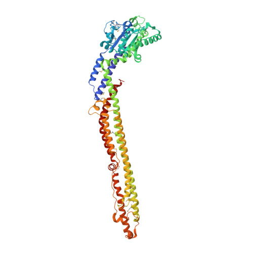
    4AUR

  • PubMed Abstract: 

    Escherichia coli (ETEC) strain H10407 contains a GTPase virulence factor, LeoA, which is encoded on a pathogenicity island and has been shown to enhance toxin release, potentially through vesicle secretion. By sequence comparisons and X-ray structure determination we now identify LeoA as a bacterial dynamin-like protein (DLP). Proteins of the dynamin family remodel membranes and were once thought to be restricted to eukaryotes. In ETEC H10407 LeoA localises to the periplasm where it forms a punctate localisation pattern. Bioinformatic analyses of leoA and the two upstream genes leoB and leoC suggest that LeoA works in concert with a second dynamin-like protein, made up of LeoB and LeoC. Disruption of the leoAB genes leads to a reduction in secretion of periplasmic Tat-GFP and outer membrane OmpA. Our data suggest a role for LeoABC dynamin-like proteins in potentiating virulence through membrane vesicle associated toxin secretion.


  • Organizational Affiliation
    • MRC Laboratory of Molecular Biology, Structural Studies Division, Cambridge, United Kingdom.

Macromolecule Content 

  • Total Structure Weight: 65.91 kDa 
  • Atom Count: 4,509 
  • Modeled Residue Count: 564 
  • Deposited Residue Count: 577 
  • Unique protein chains: 1

Macromolecules

Find similar proteins by:|  3D Structure
Entity ID: 1
MoleculeChains  Sequence LengthOrganismDetailsImage
LEOA577Escherichia coli ETEC H10407Mutation(s): 0 

Small Molecules

Ligands 1 Unique
IDChains Name / Formula / InChI Key2D Diagram3D Interactions
SO4

Query on SO4


Download:Ideal Coordinates CCD File
B [auth A]SULFATE ION
O4 S
QAOWNCQODCNURD-UHFFFAOYSA-L
Modified Residues  1 Unique
IDChains TypeFormula2D DiagramParent
MSE
Query on MSE
A
L-PEPTIDE LINKINGC5 H11 N O2 SeMET

Experimental Data & Validation

Experimental Data

  • Method: X-RAY DIFFRACTION
  • Resolution: 2.70 Å
  • R-Value Free:  0.289 (Depositor), 0.207 (DCC) 
  • R-Value Work:  0.213 (Depositor) 
  • R-Value Observed: 0.216 (Depositor) 
Space Group: C 1 2 1
Unit Cell:
Length ( Å )Angle ( ˚ )
a = 185.515α = 90
b = 53.659β = 96.65
c = 73.949γ = 90
Software Package:
Software NamePurpose
REFMACrefinement
MOSFLMdata reduction
SCALAdata scaling
SOLVEphasing

Structure Validation

View Full Validation Report


Entry History 

Deposition Data

Revision History  (Full details and data files)

  • Version 1.0: 2013-08-28
    Type: Initial release
  • Version 1.1: 2014-10-22
    Changes: Data collection, Database references
  • Version 1.2: 2024-11-20
    Changes: Data collection, Database references, Derived calculations, Other, Structure summary