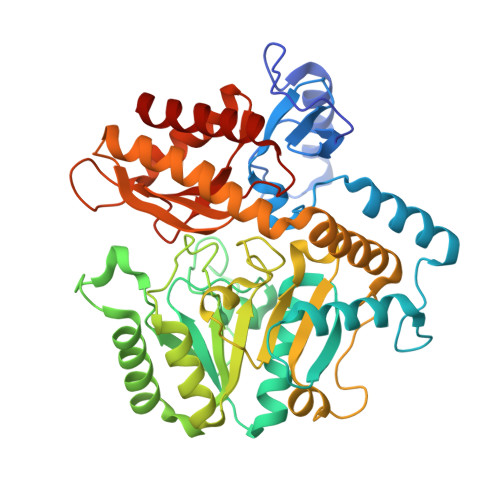
4ATQ | pdb_00004atq

GABA-transaminase A1R958 in complex with external aldimine PLP-GABA adduct


Experimental Data Snapshot

  • Method: X-RAY DIFFRACTION
  • Resolution: 2.75 Å
  • R-Value Free: 
    0.248 (Depositor), 0.240 (DCC) 
  • R-Value Work: 
    0.202 (Depositor), 0.200 (DCC) 
  • R-Value Observed: 
    0.204 (Depositor) 

Starting Model: experimental
View more details

wwPDB Validation   3D Report Full Report


Ligand Structure Quality Assessment 

Created with Raphaël 2.3.0Worse 01 BetterLigand structure goodness of fit to experimental dataBest fitted PLPClick on this verticalbar to view details

This is version 2.1 of the entry. See complete history


Literature

Structures of a Gamma-Aminobutyrate (Gaba) Transaminase from the S-Triazine-Degrading Organism Arthrobacter Aurescens Tc1 in Complex with Plp and with its External Aldimine Plp- Gaba Adduct.

Bruce, H.Nguyen Tuan, A.Mangas Sanchez, J.Leese, C.Hopwood, J.Hyde, R.Hart, S.Turkenburg, J.P.Grogan, G.

(2012) Acta Crystallogr Sect F Struct Biol Cryst Commun 68: 1175

  • DOI: https://doi.org/10.1107/S1744309112030023
  • Primary Citation of Related Structures:  
    4ATP, 4ATQ

  • PubMed Abstract: 

    Two complex structures of the γ-aminobutyrate (GABA) transaminase A1R958 from Arthrobacter aurescens TC1 are presented. The first, determined to a resolution of 2.80 Å, features the internal aldimine formed by reaction between the ℇ-amino group of Lys295 and the cofactor pyridoxal phosphate (PLP); the second, determined to a resolution of 2.75 Å, features the external aldimine adduct formed between PLP and GABA in the first half-reaction. This is the first structure of a microbial GABA transaminase in complex with its natural external aldimine and reveals the molecular determinants of GABA binding in this enzyme.


  • Organizational Affiliation

    York Structural Biology Laboratory, Department of Chemistry, University of York, Heslington, York YO10 5DD, England.


Macromolecules
Find similar proteins by:  (by identity cutoff)  |  3D Structure
Entity ID: 1
MoleculeChains Sequence LengthOrganismDetailsImage
4-AMINOBUTYRATE TRANSAMINASE
A, B, C, D, E
456Paenarthrobacter aurescens TC1Mutation(s): 0 
EC: 2.6.1.19
UniProt
Find proteins for A1R958 (Paenarthrobacter aurescens (strain TC1))
Explore A1R958 
Go to UniProtKB:  A1R958
Entity Groups  
Sequence Clusters30% Identity50% Identity70% Identity90% Identity95% Identity100% Identity
UniProt GroupA1R958
Sequence Annotations
Expand
  • Reference Sequence
Small Molecules
Ligands 2 Unique
IDChains Name / Formula / InChI Key2D Diagram3D Interactions
PLP
Query on PLP

Download Ideal Coordinates CCD File 
AA [auth H]
CA [auth I]
EA [auth J]
GA [auth K]
IA [auth L]
PYRIDOXAL-5'-PHOSPHATE
C8 H10 N O6 P
NGVDGCNFYWLIFO-UHFFFAOYSA-N
ABU
Query on ABU

Download Ideal Coordinates CCD File 
BA [auth H]
DA [auth I]
FA [auth J]
HA [auth K]
JA [auth L]
GAMMA-AMINO-BUTANOIC ACID
C4 H9 N O2
BTCSSZJGUNDROE-UHFFFAOYSA-N
Experimental Data & Validation

Experimental Data

  • Method: X-RAY DIFFRACTION
  • Resolution: 2.75 Å
  • R-Value Free:  0.248 (Depositor), 0.240 (DCC) 
  • R-Value Work:  0.202 (Depositor), 0.200 (DCC) 
  • R-Value Observed: 0.204 (Depositor) 
Space Group: P 21 21 2
Unit Cell:
Length ( Å )Angle ( ˚ )
a = 176.71α = 90
b = 292.83β = 90
c = 105.96γ = 90
Software Package:
Software NamePurpose
REFMACrefinement
xia2data scaling
MOLREPphasing

Structure Validation

View Full Validation Report


Ligand Structure Quality Assessment 

Created with Raphaël 2.3.0Worse 01 BetterLigand structure goodness of fit to experimental dataBest fitted PLPClick on this verticalbar to view details

Entry History 

Deposition Data

Revision History  (Full details and data files)

  • Version 1.0: 2012-10-10
    Type: Initial release
  • Version 1.1: 2012-10-17
    Changes: Database references
  • Version 2.0: 2023-11-15
    Changes: Atomic model, Data collection, Database references, Derived calculations, Other
  • Version 2.1: 2023-12-20
    Changes: Refinement description