4ASS

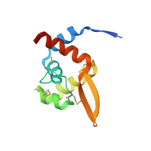
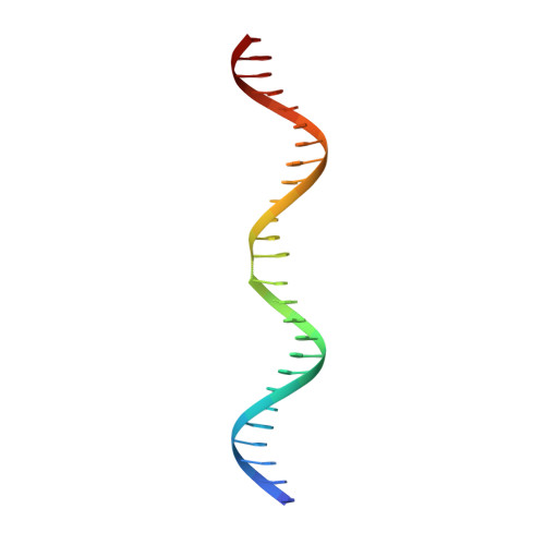
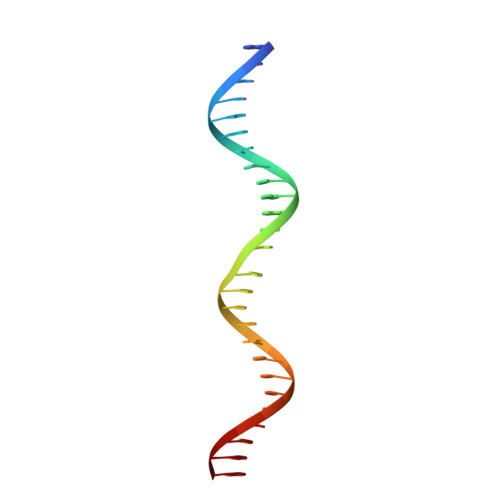
TubR bound to tubC - 26 bp - from Bacillus thuringiensis serovar israelensis pBtoxis


Experimental Data Snapshot

  • Method: X-RAY DIFFRACTION
  • Resolution: 7.00 Å
  • R-Value Free: 0.484 
  • R-Value Work: 0.408 
  • R-Value Observed: 0.412 

Starting Model: experimental
View more details

wwPDB Validation   3D Report Full Report


This is version 1.3 of the entry. See complete history


Literature

Superstructure of the Centromeric Complex of Tubzrc Plasmid Partitioning Systems.

Aylett, C.H.S.Lowe, J.

(2012) Proc Natl Acad Sci U S A 109: 16522

  • DOI: https://doi.org/10.1073/pnas.1210899109
  • Primary Citation of Related Structures:  
    4ASN, 4ASO, 4ASS

  • PubMed Abstract: 

    Bacterial plasmid partitioning systems segregate plasmids into each daughter cell. In the well-understood ParMRC plasmid partitioning system, adapter protein ParR binds to centromere parC, forming a helix around which the DNA is externally wrapped. This complex stabilizes the growth of a filament of actin-like ParM protein, which pushes the plasmids to the poles. The TubZRC plasmid partitioning system consists of two proteins, tubulin-like TubZ and TubR, and a DNA centromere, tubC, which perform analogous roles to those in ParMRC, despite being unrelated in sequence and structure. We have dissected in detail the binding sites that comprise Bacillus thuringiensis tubC, visualized the TubRC complex by electron microscopy, and determined a crystal structure of TubR bound to the tubC repeat. We show that the TubRC complex takes the form of a flexible DNA-protein filament, formed by lateral coating along the plasmid from tubC, the full length of which is required for the successful in vitro stabilization of TubZ filaments. We also show that TubR from Bacillus megaterium forms a helical superstructure resembling that of ParR. We suggest that the TubRC DNA-protein filament may bind to, and stabilize, the TubZ filament by forming such a ring-like structure around it. The helical superstructure of this TubRC may indicate convergent evolution between the actin-containing ParMRC and tubulin-containing TubZRC systems.


  • Organizational Affiliation

    Medical Research Council Laboratory of Molecular Biology, Division of Structural Studies, Cambridge, United Kingdom.


Macromolecules

Find similar proteins by:  (by identity cutoff)  |  3D Structure
Entity ID: 1
MoleculeChains Sequence LengthOrganismDetailsImage
TUBR FROM BACILLUS THURINGIENSIS PBTOXIS
A, B, C, D, E
A, B, C, D, E, F, G, H, I
104Bacillus thuringiensisMutation(s): 0 
UniProt
Find proteins for Q8KNP2 (Bacillus thuringiensis subsp. israelensis)
Explore Q8KNP2 
Go to UniProtKB:  Q8KNP2
Entity Groups  
Sequence Clusters30% Identity50% Identity70% Identity90% Identity95% Identity100% Identity
UniProt GroupQ8KNP2
Sequence Annotations
Expand
  • Reference Sequence
Find similar nucleic acids by:  (by identity cutoff)  |  3D Structure
Entity ID: 2
MoleculeChains LengthOrganismImage
TUBC FROM BACILLUS THURINGIENSIS PBTOXIS 26 BPJ [auth Y]26Bacillus thuringiensis
Sequence Annotations
Expand
  • Reference Sequence
Find similar nucleic acids by:  (by identity cutoff)  |  3D Structure
Entity ID: 3
MoleculeChains LengthOrganismImage
TUBC FROM BACILLUS THURINGIENSIS PBTOXIS 26 BPK [auth Z]26Bacillus thuringiensis
Sequence Annotations
Expand
  • Reference Sequence
Small Molecules
Modified Residues  1 Unique
IDChains TypeFormula2D DiagramParent
MSE
Query on MSE
A, B, C, D, E
A, B, C, D, E, F, G, H, I
L-PEPTIDE LINKINGC5 H11 N O2 SeMET
Experimental Data & Validation

Experimental Data

  • Method: X-RAY DIFFRACTION
  • Resolution: 7.00 Å
  • R-Value Free: 0.484 
  • R-Value Work: 0.408 
  • R-Value Observed: 0.412 
  • Space Group: I 2 2 2
Unit Cell:
Length ( Å )Angle ( ˚ )
a = 87.561α = 90
b = 162.352β = 90
c = 285.394γ = 90
Software Package:
Software NamePurpose
PHENIXrefinement
XDSdata reduction
SCALAdata scaling
PHASERphasing

Structure Validation

View Full Validation Report


Entry History 

Deposition Data

Revision History  (Full details and data files)

  • Version 1.0: 2012-10-03
    Type: Initial release
  • Version 1.1: 2012-10-24
    Changes: Database references
  • Version 1.2: 2023-12-20
    Changes: Data collection, Database references, Derived calculations, Other, Refinement description
  • Version 1.3: 2024-10-16
    Changes: Structure summary