4ARZ

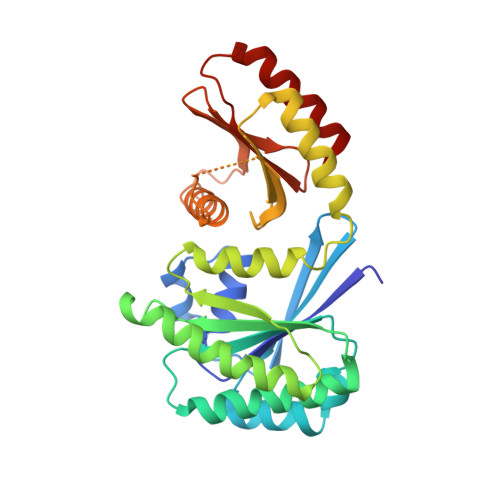
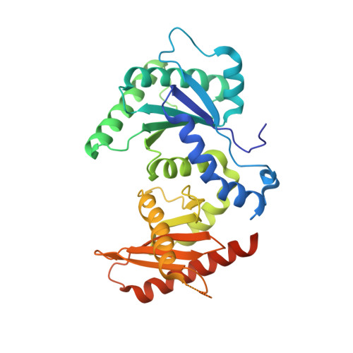
The crystal structure of Gtr1p-Gtr2p complexed with GTP-GDP


Experimental Data Snapshot

  • Method: X-RAY DIFFRACTION
  • Resolution: 3.10 Å
  • R-Value Free: 0.272 
  • R-Value Work: 0.225 
  • R-Value Observed: 0.225 

wwPDB Validation   3D Report Full Report


Ligand Structure Quality Assessment 


This is version 1.2 of the entry. See complete history


Literature

Crystal Structure of the Gtr1Pgtp-Gtr2Pgdp Complex Reveals Large Structural Rearrangements Triggered by GTP-to-Gdp Conversion

Jeong, J.H.Lee, K.H.Kim, Y.M.Kim, D.H.Oh, B.H.Kim, Y.G.

(2012) J Biol Chem 287: 29648

  • DOI: https://doi.org/10.1074/jbc.C112.384420
  • Primary Citation of Related Structures:  
    4ARZ

  • PubMed Abstract: 

    The heterodimeric Rag GTPases consisting of RagA (or RagB) and RagC (or RagD) are the key regulator activating the target of rapamycin complex 1 (TORC1) in response to the level of amino acids. The heterodimer between GTP-loaded RagA/B and GDP-loaded RagC/D is the most active form that binds Raptor and leads to the activation of TORC1. Here, we present the crystal structure of Gtr1p(GTP)-Gtr2p(GDP), the active yeast Rag GTPase heterodimer. The structure reveals that GTP-to-GDP conversion on Gtr2p results in a large conformational transition of this subunit, including a large scale rearrangement of a long segment whose corresponding region in RagA is involved in binding to Raptor. In addition, the two GTPase domains of the heterodimer are brought to contact with each other, but without causing any conformational change of the Gtr1p subunit. These features explain how the nucleotide-bound statuses of the two GTPases subunits switch the Raptor binding affinity on and off.


  • Organizational Affiliation

    Pohang Accelerator Laboratory, Pohang University of Science and Technology, Pohang, Kyungbuk 790-784, Korea.


Macromolecules
Find similar proteins by:  (by identity cutoff)  |  3D Structure
Entity ID: 1
MoleculeChains Sequence LengthOrganismDetailsImage
GTP-BINDING PROTEIN GTR1310Saccharomyces cerevisiaeMutation(s): 0 
EC: 3.6.5
UniProt
Find proteins for Q00582 (Saccharomyces cerevisiae (strain ATCC 204508 / S288c))
Explore Q00582 
Go to UniProtKB:  Q00582
Entity Groups  
Sequence Clusters30% Identity50% Identity70% Identity90% Identity95% Identity100% Identity
UniProt GroupQ00582
Sequence Annotations
Expand
  • Reference Sequence
Find similar proteins by:  (by identity cutoff)  |  3D Structure
Entity ID: 2
MoleculeChains Sequence LengthOrganismDetailsImage
GTP-BINDING PROTEIN GTR2341Saccharomyces cerevisiaeMutation(s): 0 
EC: 3.6.5
UniProt
Find proteins for P53290 (Saccharomyces cerevisiae (strain ATCC 204508 / S288c))
Explore P53290 
Go to UniProtKB:  P53290
Entity Groups  
Sequence Clusters30% Identity50% Identity70% Identity90% Identity95% Identity100% Identity
UniProt GroupP53290
Sequence Annotations
Expand
  • Reference Sequence
Experimental Data & Validation

Experimental Data

  • Method: X-RAY DIFFRACTION
  • Resolution: 3.10 Å
  • R-Value Free: 0.272 
  • R-Value Work: 0.225 
  • R-Value Observed: 0.225 
  • Space Group: C 2 2 21
Unit Cell:
Length ( Å )Angle ( ˚ )
a = 90.712α = 90
b = 148.897β = 90
c = 117.759γ = 90
Software Package:
Software NamePurpose
CNSrefinement
HKL-2000data reduction
SCALEPACKdata scaling
PHENIXphasing

Structure Validation

View Full Validation Report


Ligand Structure Quality Assessment 


Entry History 

Deposition Data

Revision History  (Full details and data files)

  • Version 1.0: 2012-07-25
    Type: Initial release
  • Version 1.1: 2012-09-05
    Changes: Database references
  • Version 1.2: 2024-05-08
    Changes: Data collection, Database references, Derived calculations, Other