4AQS | pdb_00004aqs

Laminin beta1 LN-LE1-4 structure


Experimental Data Snapshot

  • Method: X-RAY DIFFRACTION
  • Resolution: 3.11 Å
  • R-Value Free: 
    0.287 (Depositor), 0.288 (DCC) 
  • R-Value Work: 
    0.246 (Depositor) 
  • R-Value Observed: 
    0.246 (Depositor) 

Starting Models: experimental
View more details

wwPDB Validation 3D Report Full Report

Validation slider image for 4AQS

Ligand Structure Quality Assessment 


This is version 2.2 of the entry. See complete history

Literature

Crystal Structures of the Network-Forming Short-Arm Tips of the Laminin Beta1 and Gamma1 Chains.

Carafoli, F.Hussain, S.Hohenester, E.

(2012) PLoS One 7: 42473

  • DOI: https://doi.org/10.1371/journal.pone.0042473
  • Primary Citation Related Structures: 
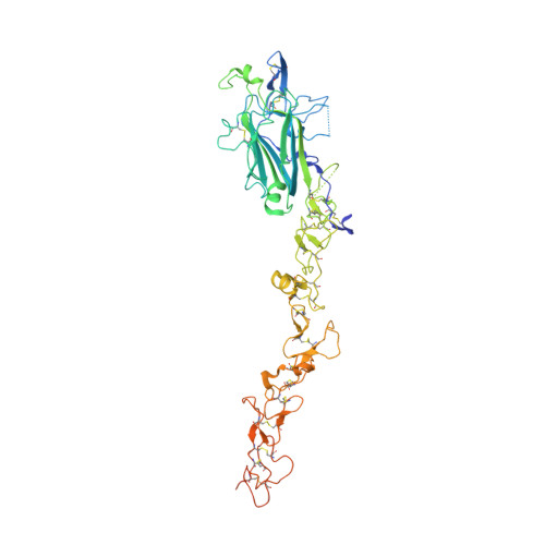
    4AQS, 4AQT

  • PubMed Abstract: 

    The heterotrimeric laminins are a defining component of basement membranes and essential for tissue formation and function in all animals. The three short arms of the cross-shaped laminin molecule are composed of one chain each and their tips mediate the formation of a polymeric network. The structural basis for laminin polymerisation is unknown. We have determined crystal structures of the short-arm tips of the mouse laminin β1 and γ1 chains, which are grossly similar to the previously determined structure of the corresponding α5 chain region. The short-arm tips consist of a laminin N-terminal (LN) domain that is attached like the head of a flower to a rod-like stem formed by tandem laminin-type epidermal growth factor-like (LE) domains. The LN domain is a β-sandwich with elaborate loop regions that differ between chains. The γ1 LN domain uniquely contains a calcium binding site. The LE domains have little regular structure and are stabilised by cysteines that are disulphide-linked 1-3, 2-4, 5-6 and 7-8 in all chains. The LN surface is not conserved across the α, β and γ chains, but within each chain subfamily there is a striking concentration of conserved residues on one face of the β-sandwich, while the opposite face invariably is shielded by glycans. We propose that the extensive conserved patches on the β and γ LN domains mediate the binding of these two chains to each other, and that the α chain LN domain subsequently binds to the composite β-γ surface. Mutations in the laminin β2 LN domain causing Pierson syndrome are likely to impair the folding of the β2 chain or its ability to form network interactions.


  • Organizational Affiliation
    • Department of Life Sciences, Imperial College London, London, United Kingdom.

Macromolecule Content 

  • Total Structure Weight: 59.82 kDa 
  • Atom Count: 3,578 
  • Modeled Residue Count: 454 
  • Deposited Residue Count: 525 
  • Unique protein chains: 1

Macromolecules

Find similar proteins by:|  3D Structure
Entity ID: 1
MoleculeChains  Sequence LengthOrganismDetailsImage
LAMININ SUBUNIT BETA-1525Mus musculusMutation(s): 0 
UniProt & NIH Common Fund Data Resources
Find proteins for P02469 (Mus musculus)
Explore P02469 
Go to UniProtKB:  P02469
IMPC:  MGI:96743
Entity Groups
Sequence Clusters30% Identity50% Identity70% Identity90% Identity95% Identity100% Identity
UniProt GroupP02469
Glycosylation
Glycosylation Sites: 2Go to GlyGen: P02469-1
Sequence Annotations
Expand
Reference Sequence

Oligosaccharides

Help  
Entity ID: 2
MoleculeChains Length2D Diagram GlycosylationD Interactions
alpha-D-mannopyranose-(1-3)-beta-D-mannopyranose-(1-4)-2-acetamido-2-deoxy-beta-D-glucopyranose-(1-4)-[beta-L-fucopyranose-(1-6)]2-acetamido-2-deoxy-beta-D-glucopyranose
B
5N-Glycosylation
Glycosylation Resources
GlyTouCan: G26551VB
GlyCosmos: G26551VB
GlyGen: G26551VB

Small Molecules

Ligands 1 Unique
IDChains Name / Formula / InChI Key2D Diagram3D Interactions
NAG

Query on NAG


Download:Ideal Coordinates CCD File
C [auth A]2-acetamido-2-deoxy-beta-D-glucopyranose
C8 H15 N O6
OVRNDRQMDRJTHS-FMDGEEDCSA-N

Experimental Data & Validation

Experimental Data

  • Method: X-RAY DIFFRACTION
  • Resolution: 3.11 Å
  • R-Value Free:  0.287 (Depositor), 0.288 (DCC) 
  • R-Value Work:  0.246 (Depositor) 
  • R-Value Observed: 0.246 (Depositor) 
Space Group: C 2 2 21
Unit Cell:
Length ( Å )Angle ( ˚ )
a = 143.99α = 90
b = 152.95β = 90
c = 92.985γ = 90
Software Package:
Software NamePurpose
CNSrefinement
iMOSFLMdata reduction
SCALAdata scaling
PHASERphasing

Structure Validation

View Full Validation Report


Ligand Structure Quality Assessment 


Entry History 

Deposition Data

Revision History  (Full details and data files)

  • Version 1.0: 2012-08-15
    Type: Initial release
  • Version 1.1: 2015-01-14
    Changes: Atomic model, Derived calculations
  • Version 2.0: 2020-07-29
    Type: Remediation
    Reason: Carbohydrate remediation
    Changes: Atomic model, Data collection, Derived calculations, Other, Structure summary
  • Version 2.1: 2023-12-20
    Changes: Data collection, Database references, Refinement description, Structure summary
  • Version 2.2: 2024-10-23
    Changes: Structure summary