4AQB

MBL-Ficolin Associated Protein-1, MAP-1 aka MAP44


Experimental Data Snapshot

  • Method: X-RAY DIFFRACTION
  • Resolution: 4.20 Å
  • R-Value Free: 0.301 
  • R-Value Work: 0.279 
  • R-Value Observed: 0.280 

Starting Models: experimental
View more details

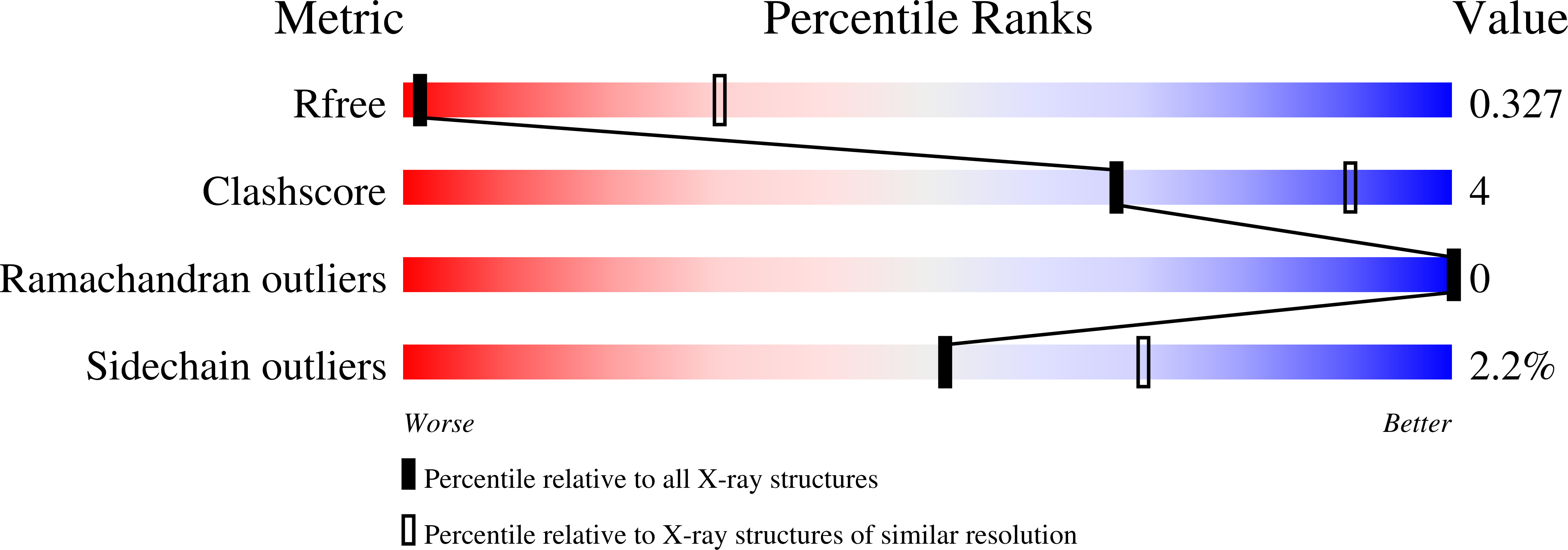
wwPDB Validation   3D Report Full Report


This is version 2.1 of the entry. See complete history


Literature

Crystal Structure and Functional Characterization of the Complement Regulator Mannose-Binding Lectin (Mbl)/Ficolin-Associated Protein-1 (Map-1).

Skjoedt, M.O.Roversi, P.Hummelshoj, T.Palarasah, Y.Rosbjerg, A.Johnson, S.Lea, S.M.Garred, P.

(2012) J Biol Chem 287: 32913

  • DOI: https://doi.org/10.1074/jbc.M112.386680
  • Primary Citation of Related Structures:  
    4AQB

  • PubMed Abstract: 

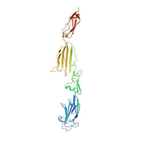
    The human lectin complement pathway activation molecules comprise mannose-binding lectin (MBL) and ficolin-1, -2, and -3 in complex with associated serine proteases MASP-1, -2, and -3 and the non-enzymatic small MBL associated protein or sMAP. Recently, a novel plasma protein named MBL/ficolin-associated protein-1 (MAP-1) was identified in humans. This protein is the result of a differential splicing of the MASP1 gene and includes the major part of the heavy chain but lacks the serine protease domain. We investigated the direct interactions of MAP-1 and MASP-3 with ficolin-3 and MBL using surface plasmon resonance and found affinities around 5 nm and 2.5 nm, respectively. We studied structural aspects of MAP-1 and could show by multi-angle laser light scattering that MAP-1 forms a calcium-dependent homodimer in solution. We were able to determine the crystal structure of MAP-1, which also contains a head-to-tail dimer ∼146 Å long. This structure of MAP-1 also enables modeling and assembly of the MASP-1 molecule in its entirety. Finally we found that MAP-1 competes with all three MASPs for ligand binding and is able to mediate a strong dose-dependent inhibitory effect on the lectin pathway activation, as measured by levels of C3 and C9.


  • Organizational Affiliation

    Laboratory of Molecular Medicine, Department of Clinical Immunology, Rigshospitalet, Faculty of Health Sciences, University of Copenhagen, DK 2100 Copenhagen, Denmark. moskjoedt@gmail.com


Macromolecules
Find similar proteins by:  (by identity cutoff)  |  3D Structure
Entity ID: 1
MoleculeChains Sequence LengthOrganismDetailsImage
MANNAN-BINDING LECTIN SERINE PROTEASE 1361Homo sapiensMutation(s): 0 
UniProt & NIH Common Fund Data Resources
Find proteins for P48740 (Homo sapiens)
Explore P48740 
Go to UniProtKB:  P48740
GTEx:  ENSG00000127241 
Entity Groups  
Sequence Clusters30% Identity50% Identity70% Identity90% Identity95% Identity100% Identity
UniProt GroupP48740
Glycosylation
Glycosylation Sites: 2Go to GlyGen: P48740-1
Sequence Annotations
Expand
  • Reference Sequence
Oligosaccharides

Help

Entity ID: 2
MoleculeChains Length2D Diagram Glycosylation3D Interactions
alpha-D-mannopyranose-(1-3)-[alpha-D-mannopyranose-(1-6)]beta-D-mannopyranose-(1-4)-2-acetamido-2-deoxy-beta-D-glucopyranose-(1-4)-2-acetamido-2-deoxy-beta-D-glucopyranose
B
5N-Glycosylation
Glycosylation Resources
GlyTouCan:  G22768VO
GlyCosmos:  G22768VO
GlyGen:  G22768VO
Entity ID: 3
MoleculeChains Length2D Diagram Glycosylation3D Interactions
alpha-D-mannopyranose-(1-3)-beta-D-mannopyranose-(1-4)-2-acetamido-2-deoxy-beta-D-glucopyranose-(1-4)-2-acetamido-2-deoxy-beta-D-glucopyranose
C
4N-Glycosylation
Glycosylation Resources
GlyTouCan:  G81315DD
GlyCosmos:  G81315DD
GlyGen:  G81315DD
Experimental Data & Validation

Experimental Data

  • Method: X-RAY DIFFRACTION
  • Resolution: 4.20 Å
  • R-Value Free: 0.301 
  • R-Value Work: 0.279 
  • R-Value Observed: 0.280 
  • Space Group: P 43 2 2
Unit Cell:
Length ( Å )Angle ( ˚ )
a = 94.15α = 90
b = 94.15β = 90
c = 242.57γ = 90
Software Package:
Software NamePurpose
BUSTERrefinement
XDSdata reduction
XSCALEdata scaling
PHASERphasing

Structure Validation

View Full Validation Report


Entry History 

Deposition Data

Revision History  (Full details and data files)

  • Version 1.0: 2012-08-08
    Type: Initial release
  • Version 1.1: 2012-10-03
    Changes: Database references
  • Version 2.0: 2020-07-29
    Type: Remediation
    Reason: Carbohydrate remediation
    Changes: Advisory, Atomic model, Data collection, Derived calculations, Other, Structure summary
  • Version 2.1: 2023-12-20
    Changes: Data collection, Database references, Refinement description, Structure summary