4APV | pdb_00004apv

The Klebsiella pneumoniae primosomal PriB protein: identification, crystal structure, and ssDNA binding mode


Experimental Data Snapshot

  • Method: X-RAY DIFFRACTION
  • Resolution: 2.10 Å
  • R-Value Free: 
    0.274 (Depositor), 0.276 (DCC) 
  • R-Value Work: 
    0.198 (Depositor), 0.199 (DCC) 
  • R-Value Observed: 
    0.205 (Depositor) 

Starting Model: experimental
View more details

wwPDB Validation 3D Report Full Report

Validation slider image for 4APV

This is version 1.3 of the entry. See complete history

Literature

Crystal Structure and DNA-Binding Mode of Klebsiella Pneumoniae Primosomal Prib Protein.

Huang, Y.Lo, Y.H.Huang, W.Huang, C.Y.

(2012) Genes Cells 17: 837

  • DOI: https://doi.org/10.1111/gtc.12001
  • Primary Citation Related Structures: 
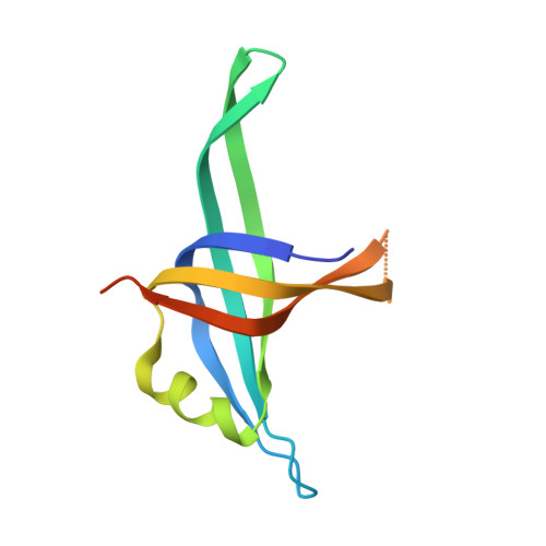
    4APV

  • PubMed Abstract: 

    PriB is a primosomal DNA replication protein required for the re-initiation of replication in bacteria. In this study, we investigated the gene expression of PriB in Klebsiella pneumoniae (KpPriB) and characterized the gene product through crystal structural and functional analyses. Quantitative polymerase chain reaction analysis (Q-PCR) indicated that the 104-aa priB was expressed in K. pneumoniae with a C(T) value of 22.4. The crystal structure of KpPriB (Protein Data Bank entry: 4APV) determined at a resolution of 2.1 Å was similar to that of Escherichia coli PriB (EcPriB). KpPriB formed a single complex with single-stranded DNA (ssDNA) of different lengths, suggesting a highly cooperative process. Structure-based mutational analysis revealed that substitution at K18, F42, R44, W47, K82, K84, or K89 but not R34 in KpPriB had a significant effect on both ssDNA and double-stranded DNA (dsDNA) binding. Based on these findings, the known ssDNA interaction sites of PriB were expanded to include R44 and F42, thus allowing nucleic acids to wrap around the whole PriB protein.


  • Organizational Affiliation
    • Department of Biomedical Sciences, Chung Shan Medical University, No. 110, Sec. 1, Chien-Kuo N. Rd, Taichung City, Taiwan.

Macromolecule Content 

  • Total Structure Weight: 12.56 kDa 
  • Atom Count: 765 
  • Modeled Residue Count: 98 
  • Deposited Residue Count: 112 
  • Unique protein chains: 1

Macromolecules

Find similar proteins by:|  3D Structure
Entity ID: 1
MoleculeChains  Sequence LengthOrganismDetailsImage
PRIMOSOMAL REPLICATION PROTEIN N112Klebsiella pneumoniaeMutation(s): 0 

Experimental Data & Validation

Experimental Data

  • Method: X-RAY DIFFRACTION
  • Resolution: 2.10 Å
  • R-Value Free:  0.274 (Depositor), 0.276 (DCC) 
  • R-Value Work:  0.198 (Depositor), 0.199 (DCC) 
  • R-Value Observed: 0.205 (Depositor) 
Space Group: C 1 2 1
Unit Cell:
Length ( Å )Angle ( ˚ )
a = 64.773α = 90
b = 36.934β = 113.77
c = 39.451γ = 90
Software Package:
Software NamePurpose
PHENIXrefinement
HKL-2000data reduction
HKL-2000data scaling
PHASERphasing

Structure Validation

View Full Validation Report


Entry History 

Deposition Data

Revision History  (Full details and data files)

  • Version 1.0: 2012-04-25
    Type: Initial release
  • Version 1.1: 2013-01-23
    Changes: Database references
  • Version 1.2: 2023-12-20
    Changes: Data collection, Database references, Other, Refinement description
  • Version 1.3: 2024-11-20
    Changes: Structure summary