4APH | pdb_00004aph

Human angiotensin-converting enzyme in complex with angiotensin-II


Experimental Data Snapshot

  • Method: X-RAY DIFFRACTION
  • Resolution: 1.99 Å
  • R-Value Free: 
    0.240 (Depositor), 0.241 (DCC) 
  • R-Value Work: 
    0.194 (Depositor), 0.194 (DCC) 
  • R-Value Observed: 
    0.196 (Depositor) 

Starting Model: experimental
View more details

wwPDB Validation 3D Report Full Report

Validation slider image for 4APH

Ligand Structure Quality Assessment 


This is version 3.0 of the entry. See complete history

Literature

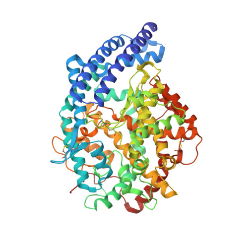
Molecular Recognition and Regulation of Human Angiotensin-I Converting Enzyme (Ace) Activity by Natural Inhibitory Peptides.

Masuyer, G.Schwager, S.L.U.Sturrock, E.D.Isaac, R.E.Acharya, K.R.

(2012) Sci Rep 2: 717

  • DOI: https://doi.org/10.1038/srep00717
  • Primary Citation Related Structures: 
    4APH, 4APJ

  • PubMed Abstract: 

    Angiotensin-I converting enzyme (ACE), a two-domain dipeptidylcarboxypeptidase, is a key regulator of blood pressure as a result of its critical role in the renin-angiotensin-aldosterone and kallikrein-kinin systems. Hence it is an important drug target in the treatment of cardiovascular diseases. ACE is primarily known for its ability to cleave angiotensin I (Ang I) to the vasoactive octapeptide angiotensin II (Ang II), but is also able to cleave a number of other substrates including the vasodilator bradykinin and N-acetyl-Ser-Asp-Lys-Pro (Ac-SDKP), a physiological modulator of hematopoiesis. For the first time we provide a detailed biochemical and structural basis for the domain selectivity of the natural peptide inhibitors of ACE, bradykinin potentiating peptide b and Ang II. Moreover, Ang II showed selective competitive inhibition of the carboxy-terminal domain of human somatic ACE providing evidence for a regulatory role in the human renin-angiotensin system (RAS).


  • Organizational Affiliation
    • Department of Biology and Biochemistry, University of Bath, Claverton Down, Bath, UK.

Macromolecule Content 

  • Total Structure Weight: 70.71 kDa 
  • Atom Count: 5,319 
  • Modeled Residue Count: 590 
  • Deposited Residue Count: 597 
  • Unique protein chains: 2

Macromolecules

Find similar proteins by:|  3D Structure
Entity ID: 1
MoleculeChains  Sequence LengthOrganismDetailsImage
ANGIOTENSIN-CONVERTING ENZYME589Homo sapiensMutation(s): 0 
EC: 3.2.1 (PDB Primary Data), 3.4.15.1 (PDB Primary Data)
UniProt & NIH Common Fund Data Resources
Find proteins for P12821 (Homo sapiens)
Explore P12821 
Go to UniProtKB:  P12821
PHAROS:  P12821
GTEx:  ENSG00000159640 
Entity Groups
Sequence Clusters30% Identity50% Identity70% Identity90% Identity95% Identity100% Identity
UniProt GroupP12821
Glycosylation
Glycosylation Sites: 2Go to GlyGen: P12821-1
Sequence Annotations
Expand
Reference Sequence
Find similar proteins by:  Sequence   |   3D Structure  
Entity ID: 2
MoleculeChains  Sequence LengthOrganismDetailsImage
ANGIOTENSIN-2B [auth P]8Homo sapiensMutation(s): 0 
UniProt & NIH Common Fund Data Resources
Find proteins for P01019 (Homo sapiens)
Explore P01019 
Go to UniProtKB:  P01019
PHAROS:  P01019
GTEx:  ENSG00000135744 
Entity Groups
UniProt GroupP01019
Sequence Annotations
Expand
Reference Sequence

Oligosaccharides

Help  
Entity ID: 3
MoleculeChains Length2D Diagram GlycosylationD Interactions
beta-D-mannopyranose-(1-4)-2-acetamido-2-deoxy-beta-D-glucopyranose-(1-4)-2-acetamido-2-deoxy-beta-D-glucopyranoseC [auth B]3N-Glycosylation
Glycosylation Resources
GlyTouCan: G15407YE
GlyCosmos: G15407YE
GlyGen: G15407YE

Small Molecules

Ligands 5 Unique
IDChains Name / Formula / InChI Key2D Diagram3D Interactions
PE4

Query on PE4


Download:Ideal Coordinates CCD File
G [auth A]2-{2-[2-(2-{2-[2-(2-ETHOXY-ETHOXY)-ETHOXY]-ETHOXY}-ETHOXY)-ETHOXY]-ETHOXY}-ETHANOL
C16 H34 O8
PJWQOENWHPEPKI-UHFFFAOYSA-N
NAG

Query on NAG


Download:Ideal Coordinates CCD File
H [auth A]2-acetamido-2-deoxy-beta-D-glucopyranose
C8 H15 N O6
OVRNDRQMDRJTHS-FMDGEEDCSA-N
ZN

Query on ZN


Download:Ideal Coordinates CCD File
E [auth A]ZINC ION
Zn
PTFCDOFLOPIGGS-UHFFFAOYSA-N
ACT

Query on ACT


Download:Ideal Coordinates CCD File
I [auth A],
J [auth A],
K [auth A],
L [auth A],
M [auth A]
ACETATE ION
C2 H3 O2
QTBSBXVTEAMEQO-UHFFFAOYSA-M
CL

Query on CL


Download:Ideal Coordinates CCD File
D [auth A],
F [auth A]
CHLORIDE ION
Cl
VEXZGXHMUGYJMC-UHFFFAOYSA-M

Experimental Data & Validation

Experimental Data

  • Method: X-RAY DIFFRACTION
  • Resolution: 1.99 Å
  • R-Value Free:  0.240 (Depositor), 0.241 (DCC) 
  • R-Value Work:  0.194 (Depositor), 0.194 (DCC) 
  • R-Value Observed: 0.196 (Depositor) 
Space Group: P 21 21 21
Unit Cell:
Length ( Å )Angle ( ˚ )
a = 56.5α = 90
b = 84.66β = 90
c = 133.97γ = 90
Software Package:
Software NamePurpose
REFMACrefinement
MOSFLMdata reduction
SCALAdata scaling
PHASERphasing

Structure Validation

View Full Validation Report


Ligand Structure Quality Assessment 


Entry History 

Deposition Data

Revision History  (Full details and data files)

  • Version 1.0: 2012-10-17
    Type: Initial release
  • Version 1.1: 2012-10-24
    Changes: Database references
  • Version 1.2: 2017-01-25
    Changes: Atomic model, Derived calculations, Non-polymer description, Other
  • Version 2.0: 2020-07-29
    Type: Remediation
    Reason: Carbohydrate remediation
    Changes: Advisory, Atomic model, Data collection, Derived calculations, Other, Structure summary
  • Version 2.1: 2023-12-20
    Changes: Data collection, Database references, Refinement description, Structure summary
  • Version 3.0: 2025-10-01
    Changes: Advisory, Atomic model, Data collection, Derived calculations, Structure summary