4ANM | pdb_00004anm

Complex of CK2 with a CDC7 inhibitor


Experimental Data Snapshot

  • Method: X-RAY DIFFRACTION
  • Resolution: 1.70 Å
  • R-Value Free: 
    0.202 (Depositor), 0.201 (DCC) 
  • R-Value Work: 
    0.164 (Depositor), 0.163 (DCC) 
  • R-Value Observed: 
    0.166 (Depositor) 

Starting Model: other
View more details

wwPDB Validation 3D Report Full Report

Validation slider image for 4ANM

Ligand Structure Quality Assessment 


This is version 1.1 of the entry. See complete history

Literature

Discovery of Xl413, a Potent and Selective Cdc7 Inhibitor.

Koltun, E.S.Tsuhako, A.L.Brown, D.S.Aay, N.Arcalas, A.Chan, V.Du, H.Engst, S.Ferguson, K.Franzini, M.Galan, A.Holst, C.R.Huang, P.Kane, B.Kim, M.H.Li, J.Markby, D.Mohan, M.Noson, K.Plonowski, A.Richards, S.J.Robertson, S.Shaw, K.Stott, G.Stout, T.J.Young, J.Yu, P.Zaharia, C.A.Zhang, W.Zhou, P.Nuss, J.M.Xu, W.Kearney, P.C.

(2012) Bioorg Med Chem Lett 22: 3727

  • DOI: https://doi.org/10.1016/j.bmcl.2012.04.024
  • Primary Citation Related Structures: 
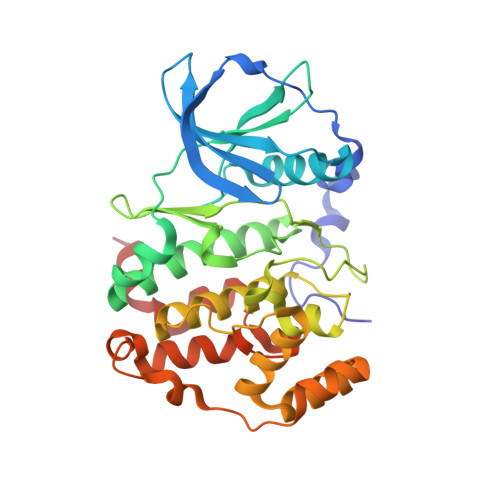
    4ANM

  • PubMed Abstract: 

    CDC7 is a serine/threonine kinase that has been shown to be required for the initiation and maintenance of DNA replication. Up-regulation of CDC7 is detected in multiple tumor cell lines, with inhibition of CDC7 resulting in cell cycle arrest. In this paper, we disclose the discovery of a potent and selective CDC7 inhibitor, XL413 (14), which was advanced into Phase 1 clinical trials. Starting from advanced lead 3, described in a preceding communication, we optimized the CDC7 potency and selectivity to demonstrate in vitro CDC7 dependent cell cycle arrest and in vivo tumor growth inhibition in a Colo-205 xenograft model.


  • Organizational Affiliation
    • Exelixis, Department of Drug Discovery, South San Francisco, CA 94080, USA. elena@numerate.com

Macromolecule Content 

  • Total Structure Weight: 39.94 kDa 
  • Atom Count: 3,125 
  • Modeled Residue Count: 328 
  • Deposited Residue Count: 335 
  • Unique protein chains: 1

Macromolecules

Find similar proteins by:|  3D Structure
Entity ID: 1
MoleculeChains  Sequence LengthOrganismDetailsImage
CASEIN KINASE II SUBUNIT ALPHA335Zea maysMutation(s): 0 
EC: 2.7.11.1
UniProt
Find proteins for P28523 (Zea mays)
Explore P28523 
Go to UniProtKB:  P28523
Entity Groups
Sequence Clusters30% Identity50% Identity70% Identity90% Identity95% Identity100% Identity
UniProt GroupP28523
Sequence Annotations
Expand
Reference Sequence

Small Molecules

Ligands 1 Unique
IDChains Name / Formula / InChI Key2D Diagram3D Interactions
WUL

Query on WUL


Download:Ideal Coordinates CCD File
B [auth A]8-BROMANYL-2-[[(3S)-3-OXIDANYLPYRROLIDIN-1-YL]METHYL]-3H-[1]BENZOFURO[3,2-D]PYRIMIDIN-4-ONE
C15 H14 Br N3 O3
FRRLLRQAGKSVMU-VIFPVBQESA-N

Experimental Data & Validation

Experimental Data

  • Method: X-RAY DIFFRACTION
  • Resolution: 1.70 Å
  • R-Value Free:  0.202 (Depositor), 0.201 (DCC) 
  • R-Value Work:  0.164 (Depositor), 0.163 (DCC) 
  • R-Value Observed: 0.166 (Depositor) 
Space Group: C 1 2 1
Unit Cell:
Length ( Å )Angle ( ˚ )
a = 142.061α = 90
b = 60.549β = 102.92
c = 44.747γ = 90
Software Package:
Software NamePurpose
REFMACrefinement
XDSdata reduction
XSCALEdata scaling
PHASERphasing

Structure Validation

View Full Validation Report


Ligand Structure Quality Assessment 


Entry History 

Deposition Data

  • Released Date: 2012-05-23 
  • Deposition Author(s): Stout, T.J.

Revision History  (Full details and data files)

  • Version 1.0: 2012-05-23
    Type: Initial release
  • Version 1.1: 2024-05-01
    Changes: Data collection, Database references, Derived calculations, Other, Refinement description