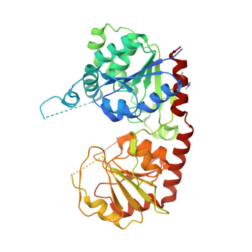
Crystal Structure of the Glycosyltransferase Snogd from the Biosynthetic Pathway of the Nogalamycin in Streptomyces Nogalater.
Claesson, M., Siitonen, V., Dobritzsch, D., Metsa-Ketela, M., Schneider, G.(2012) FEBS J 279: 3251
- PubMed: 22804797
- DOI: https://doi.org/10.1111/j.1742-4658.2012.08711.x
- Primary Citation of Related Structures:
4AMB, 4AMG, 4AN4 - PubMed Abstract:
The glycosyltransferase SnogD from Streptomyces nogalater transfers a nogalamine moiety to the metabolic intermediate 3',4'-demethoxynogalose-1-hydroxynogalamycinone during the final steps of biosynthesis of the aromatic polyketide nogalamycin. The crystal structure of recombinant SnogD, as an apo-enzyme and with a bound nucleotide, 2-deoxyuridine-5'-diphosphate, was determined to 2.6 Å resolution. Reductive methylation of SnogD was crucial for reproducible preparation of diffraction quality crystals due to creation of an additional intermolecular salt bridge between methylated lysine residue Lys384 and Glu374* from an adjacent molecule in the crystal lattice. SnogD is a dimer both in solution and in the crystal, and the enzyme subunit displays a fold characteristic of the GT-B family of glycosyltransferases. Binding of the nucleotide is associated with rearrangement of two active-site loops. Site-directed mutagenesis shows that two active-site histidine residues, His25 and His301, are critical for the glycosyltransferase activities of SnogD both in vivo and in vitro. The crystal structures and the functional data are consistent with a role for His301 in binding of the diphosphate group of the sugar donor substrate, and a function of His25 as a catalytic base in the glycosyl transfer reaction.
- Department of Medical Biochemistry and Biophysics, Karolinska Institutet, Stockholm, Sweden.
Organizational Affiliation:


