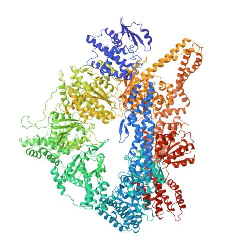
4AKI

Dynein Motor Domain - LuAc derivative


Experimental Data Snapshot

  • Method: X-RAY DIFFRACTION
  • Resolution: 3.70 Å
  • R-Value Free: 0.289 
  • R-Value Work: 0.231 
  • R-Value Observed: 0.234 

wwPDB Validation   3D Report Full Report


Ligand Structure Quality Assessment 


This is version 1.4 of the entry. See complete history


Literature

Insights Into Dynein Motor Domain Function from a 3.3 Angstrom Crystal Structure

Schmidt, H.Gleave, E.S.Carter, A.P.

(2012) Nat Struct Mol Biol 19: 492

  • DOI: https://doi.org/10.1038/nsmb.2272
  • Primary Citation of Related Structures:  
    4AI6, 4AKG, 4AKH, 4AKI

  • PubMed Abstract: 

    Dyneins power the beating of cilia and flagella, transport various intracellular cargos and are necessary for mitosis. All dyneins have a ∼300-kDa motor domain consisting of a ring of six AAA+ domains. ATP hydrolysis in the AAA+ ring drives the cyclic relocation of a motile element, the linker domain, to generate the force necessary for movement. How the linker interacts with the ring during the ATP hydrolysis cycle is not known. Here we present a 3.3-Å crystal structure of the motor domain of Saccharomyces cerevisiae cytoplasmic dynein, crystallized in the absence of nucleotides. The linker is docked to a conserved site on AAA5, which is confirmed by mutagenesis as functionally necessary. Nucleotide soaking experiments show that the main ATP hydrolysis site in dynein (AAA1) is in a low-nucleotide affinity conformation and reveal the nucleotide interactions of the other three sites (AAA2, AAA3 and AAA4).


  • Organizational Affiliation

    Medical Research Council Laboratory of Molecular Biology, Cambridge, UK.


Macromolecules
Find similar proteins by:  (by identity cutoff)  |  3D Structure
Entity ID: 1
MoleculeChains Sequence LengthOrganismDetailsImage
GLUTATHIONE S-TRANSFERASE CLASS-MU 26 KDA ISOZYME, DYNEIN HEAVY CHAIN CYTOPLASMIC
A, B
2,695Schistosoma japonicumSaccharomyces cerevisiaeMutation(s): 0 
EC: 2.5.1.18
UniProt
Find proteins for P08515 (Schistosoma japonicum)
Explore P08515 
Go to UniProtKB:  P08515
Find proteins for P36022 (Saccharomyces cerevisiae (strain ATCC 204508 / S288c))
Explore P36022 
Go to UniProtKB:  P36022
Entity Groups  
Sequence Clusters30% Identity50% Identity70% Identity90% Identity95% Identity100% Identity
UniProt GroupsP36022P08515
Sequence Annotations
Expand
  • Reference Sequence
Small Molecules
Ligands 3 Unique
IDChains Name / Formula / InChI Key2D Diagram3D Interactions
ATP
Query on ATP

Download Ideal Coordinates CCD File 
C [auth A],
H [auth B]
ADENOSINE-5'-TRIPHOSPHATE
C10 H16 N5 O13 P3
ZKHQWZAMYRWXGA-KQYNXXCUSA-N
SO4
Query on SO4

Download Ideal Coordinates CCD File 
D [auth A]
E [auth A]
F [auth A]
I [auth B]
J [auth B]
D [auth A],
E [auth A],
F [auth A],
I [auth B],
J [auth B],
K [auth B]
SULFATE ION
O4 S
QAOWNCQODCNURD-UHFFFAOYSA-L
MG
Query on MG

Download Ideal Coordinates CCD File 
G [auth A],
L [auth B]
MAGNESIUM ION
Mg
JLVVSXFLKOJNIY-UHFFFAOYSA-N
Experimental Data & Validation

Experimental Data

  • Method: X-RAY DIFFRACTION
  • Resolution: 3.70 Å
  • R-Value Free: 0.289 
  • R-Value Work: 0.231 
  • R-Value Observed: 0.234 
  • Space Group: P 1 21 1
Unit Cell:
Length ( Å )Angle ( ˚ )
a = 175.769α = 90
b = 118.186β = 90.91
c = 202.679γ = 90
Software Package:
Software NamePurpose
REFMACrefinement
MOSFLMdata reduction
Aimlessdata scaling

Structure Validation

View Full Validation Report


Ligand Structure Quality Assessment 


Entry History 

Deposition Data

Revision History  (Full details and data files)

  • Version 1.0: 2012-03-14
    Type: Initial release
  • Version 1.1: 2012-05-16
    Changes: Other
  • Version 1.2: 2015-10-21
    Changes: Source and taxonomy
  • Version 1.3: 2017-03-15
    Changes: Source and taxonomy
  • Version 1.4: 2024-05-08
    Changes: Data collection, Database references, Derived calculations, Other