4AC7 | pdb_00004ac7

The crystal structure of Sporosarcina pasteurii urease in complex with citrate


Experimental Data Snapshot

  • Method: X-RAY DIFFRACTION
  • Resolution: 1.50 Å
  • R-Value Free: 
    0.146 (Depositor), 0.156 (DCC) 
  • R-Value Work: 
    0.117 (Depositor), 0.130 (DCC) 
  • R-Value Observed: 
    0.118 (Depositor) 

Starting Model: experimental
View more details

wwPDB Validation 3D Report Full Report

Validation slider image for 4AC7

This is version 1.4 of the entry. See complete history

Literature

The Crystal Structure of Sporosarcina Pasteurii Urease in a Complex with Citrate Provides New Hints for Inhibitor Design.

Benini, S.Kosikowska, P.Cianci, M.Mazzei, L.Vara, A.G.Berlicki, L.Ciurli, S.

(2013) J Biol Inorg Chem 18: 391

  • DOI: https://doi.org/10.1007/s00775-013-0983-7
  • Primary Citation Related Structures: 
    4AC7

  • PubMed Abstract: 

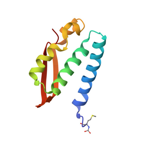
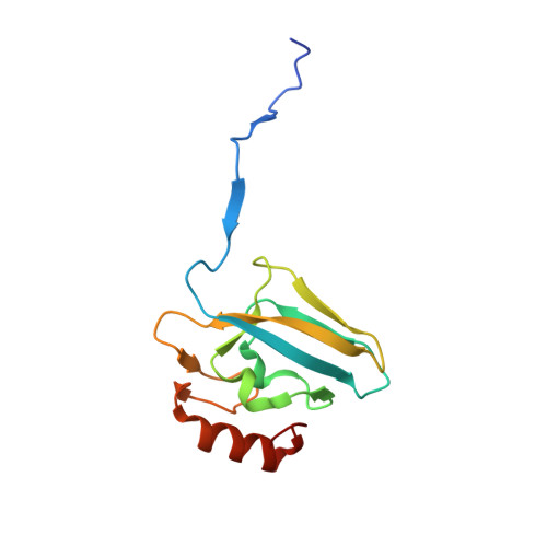
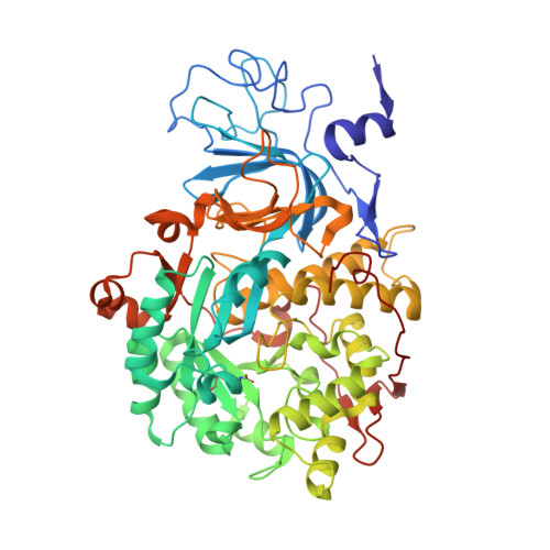
    Urease, the enzyme that catalyses the hydrolysis of urea, is a virulence factor for a large number of ureolytic bacterial human pathogens. The increasing resistance of these pathogens to common antibiotics as well as the need to control urease activity to improve the yield of soil nitrogen fertilization in agricultural applications has stimulated the development of novel classes of molecules that target urease as enzyme inhibitors. We report on the crystal structure at 1.50-Å resolution of a complex formed between citrate and urease from Sporosarcina pasteurii, a widespread and highly ureolytic soil bacterium. The fit of the ligand to the active site involves stabilizing interactions, such as a carboxylate group that binds the nickel ions at the active site and several hydrogen bonds with the surrounding residues. The citrate ligand has a significantly extended structure compared with previously reported ligands co-crystallized with urease and thus represents a unique and promising scaffold for the design of new, highly active, stable, selective inhibitors.


  • Organizational Affiliation
    • Faculty of Science and Technology, Free University of Bolzano, Bolzano, Italy.

Macromolecule Content 

  • Total Structure Weight: 88.16 kDa 
  • Atom Count: 6,830 
  • Modeled Residue Count: 792 
  • Deposited Residue Count: 796 
  • Unique protein chains: 3

Macromolecules

Find similar proteins by:|  3D Structure
Entity ID: 1
MoleculeChains  Sequence LengthOrganismDetailsImage
UREASE SUBUNIT GAMMA100Sporosarcina pasteuriiMutation(s): 0 
EC: 3.5.1.5
UniProt
Find proteins for P41022 (Sporosarcina pasteurii)
Explore P41022 
Go to UniProtKB:  P41022
Entity Groups
Sequence Clusters30% Identity50% Identity70% Identity90% Identity95% Identity100% Identity
UniProt GroupP41022
Sequence Annotations
Expand
Reference Sequence
Find similar proteins by:|  3D Structure
Entity ID: 2
MoleculeChains  Sequence LengthOrganismDetailsImage
UREASE SUBUNIT BETA126Sporosarcina pasteuriiMutation(s): 0 
EC: 3.5.1.5
UniProt
Find proteins for P41021 (Sporosarcina pasteurii)
Explore P41021 
Go to UniProtKB:  P41021
Entity Groups
Sequence Clusters30% Identity50% Identity70% Identity90% Identity95% Identity100% Identity
UniProt GroupP41021
Sequence Annotations
Expand
Reference Sequence
Find similar proteins by:|  3D Structure
Entity ID: 3
MoleculeChains  Sequence LengthOrganismDetailsImage
UREASE SUBUNIT ALPHA570Sporosarcina pasteuriiMutation(s): 0 
EC: 3.5.1.5
UniProt
Find proteins for P41020 (Sporosarcina pasteurii)
Explore P41020 
Go to UniProtKB:  P41020
Entity Groups
Sequence Clusters30% Identity50% Identity70% Identity90% Identity95% Identity100% Identity
UniProt GroupP41020
Sequence Annotations
Expand
Reference Sequence

Small Molecules

Ligands 5 Unique
IDChains Name / Formula / InChI Key2D Diagram3D Interactions
FLC

Query on FLC


Download:Ideal Coordinates CCD File
L [auth C]CITRATE ANION
C6 H5 O7
KRKNYBCHXYNGOX-UHFFFAOYSA-K
SO4

Query on SO4


Download:Ideal Coordinates CCD File
H [auth B],
S [auth C],
T [auth C],
U [auth C]
SULFATE ION
O4 S
QAOWNCQODCNURD-UHFFFAOYSA-L
EDO

Query on EDO


Download:Ideal Coordinates CCD File
D [auth A]
E [auth A]
F [auth A]
G [auth B]
M [auth C]
D [auth A],
E [auth A],
F [auth A],
G [auth B],
M [auth C],
N [auth C],
O [auth C],
P [auth C],
Q [auth C],
R [auth C]
1,2-ETHANEDIOL
C2 H6 O2
LYCAIKOWRPUZTN-UHFFFAOYSA-N
NI

Query on NI


Download:Ideal Coordinates CCD File
I [auth C],
J [auth C]
NICKEL (II) ION
Ni
VEQPNABPJHWNSG-UHFFFAOYSA-N
OH

Query on OH


Download:Ideal Coordinates CCD File
K [auth C]HYDROXIDE ION
H O
XLYOFNOQVPJJNP-UHFFFAOYSA-M
Modified Residues  2 Unique
IDChains TypeFormula2D DiagramParent
CXM
Query on CXM
A
L-PEPTIDE LINKINGC6 H11 N O4 SMET
KCX
Query on KCX
C
L-PEPTIDE LINKINGC7 H14 N2 O4LYS

Experimental Data & Validation

Experimental Data

  • Method: X-RAY DIFFRACTION
  • Resolution: 1.50 Å
  • R-Value Free:  0.146 (Depositor), 0.156 (DCC) 
  • R-Value Work:  0.117 (Depositor), 0.130 (DCC) 
  • R-Value Observed: 0.118 (Depositor) 
Space Group: P 63 2 2
Unit Cell:
Length ( Å )Angle ( ˚ )
a = 131.555α = 90
b = 131.555β = 90
c = 189.521γ = 120
Software Package:
Software NamePurpose
REFMACrefinement
XDSdata reduction
SCALAdata scaling
REFMACphasing

Structure Validation

View Full Validation Report


Entry History 

Deposition Data

Revision History  (Full details and data files)

  • Version 1.0: 2013-01-16
    Type: Initial release
  • Version 1.1: 2013-05-22
    Changes: Database references
  • Version 1.2: 2014-12-03
    Changes: Data collection
  • Version 1.3: 2018-03-14
    Changes: Data collection
  • Version 1.4: 2023-12-20
    Changes: Data collection, Database references, Derived calculations, Other, Refinement description