4AC1 | pdb_00004ac1

The structure of a fungal endo-beta-N-acetylglucosaminidase from glycosyl hydrolase family 18, at 1.3A resolution


Experimental Data Snapshot

  • Method: X-RAY DIFFRACTION
  • Resolution: 1.30 Å
  • R-Value Free: 
    0.202 (Depositor), 0.199 (DCC) 
  • R-Value Work: 
    0.184 (Depositor), 0.182 (DCC) 
  • R-Value Observed: 
    0.185 (Depositor) 

wwPDB Validation 3D Report Full Report

Validation slider image for 4AC1

Ligand Structure Quality Assessment 


This is version 1.3 of the entry. See complete history

Literature

High Resolution Crystal Structure of the Endo-N-Acetyl-Beta- D-Glucosaminidase Responsible for the Deglycosylation of Hypocrea Jecorina Cellulases.

Stals, I.Karkehabadi, S.Kim, S.Ward, M.Van Landschoot, A.Devreese, B.Sandgren, M.

(2012) PLoS One 7: 40854

  • DOI: https://doi.org/10.1371/journal.pone.0040854
  • Primary Citation Related Structures: 
    4AC1

  • PubMed Abstract: 

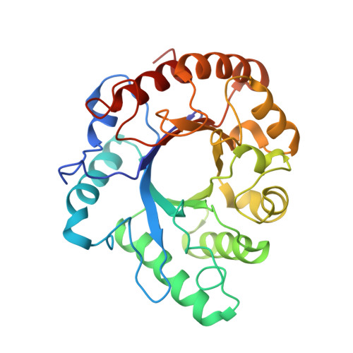
    Endo-N-acetyl-β-D-glucosaminidases (ENGases) hydrolyze the glycosidic linkage between the two N-acetylglucosamine units that make up the chitobiose core of N-glycans. The endo-N-acetyl-β-D-glucosaminidases classified into glycoside hydrolase family 18 are small, bacterial proteins with different substrate specificities. Recently two eukaryotic family 18 deglycosylating enzymes have been identified. Here, the expression, purification and the 1.3Å resolution structure of the ENGase (Endo T) from the mesophilic fungus Hypocrea jecorina (anamorph Trichoderma reesei) are reported. Although the mature protein is C-terminally processed with removal of a 46 amino acid peptide, the protein has a complete (β/α)8 TIM-barrel topology. In the active site, the proton donor (E131) and the residue stabilizing the transition state (D129) in the substrate assisted catalysis mechanism are found in almost identical positions as in the bacterial GH18 ENGases: Endo H, Endo F1, Endo F3, and Endo BT. However, the loops defining the substrate-binding cleft vary greatly from the previously known ENGase structures, and the structures also differ in some of the α-helices forming the barrel. This could reflect the variation in substrate specificity between the five enzymes. This is the first three-dimensional structure of a eukaryotic endo-N-acetyl-β-D-glucosaminidase from glycoside hydrolase family 18. A glycosylation analysis of the cellulases secreted by a Hypocrea jecorina Endo T knock-out strain shows the in vivo function of the protein. A homology search and phylogenetic analysis show that the two known enzymes and their homologues form a large but separate cluster in subgroup B of the fungal chitinases. Therefore the future use of a uniform nomenclature is proposed.


  • Organizational Affiliation
    • Faculty of Applied Bioscience Engineering, University College Ghent, Ghent, Belgium.

Macromolecule Content 

  • Total Structure Weight: 32.08 kDa 
  • Atom Count: 2,700 
  • Modeled Residue Count: 283 
  • Deposited Residue Count: 283 
  • Unique protein chains: 1

Macromolecules

Find similar proteins by:|  3D Structure
Entity ID: 1
MoleculeChains  Sequence LengthOrganismDetailsImage
ENDO-N-ACETYL-BETA-D-GLUCOSAMINIDASEA [auth X]283Trichoderma reeseiMutation(s): 0 
EC: 3.2.1.96
UniProt
Find proteins for C4RA89 (Hypocrea jecorina)
Explore C4RA89 
Go to UniProtKB:  C4RA89
Entity Groups
Sequence Clusters30% Identity50% Identity70% Identity90% Identity95% Identity100% Identity
UniProt GroupC4RA89
Glycosylation
Glycosylation Sites: 2
Sequence Annotations
Expand
Reference Sequence

Small Molecules

Ligands 3 Unique
IDChains Name / Formula / InChI Key2D Diagram3D Interactions
NAG

Query on NAG


Download:Ideal Coordinates CCD File
B [auth X],
C [auth X]
2-acetamido-2-deoxy-beta-D-glucopyranose
C8 H15 N O6
OVRNDRQMDRJTHS-FMDGEEDCSA-N
ZN

Query on ZN


Download:Ideal Coordinates CCD File
G [auth X]
H [auth X]
I [auth X]
J [auth X]
K [auth X]
G [auth X],
H [auth X],
I [auth X],
J [auth X],
K [auth X],
L [auth X],
M [auth X],
N [auth X]
ZINC ION
Zn
PTFCDOFLOPIGGS-UHFFFAOYSA-N
ACT

Query on ACT


Download:Ideal Coordinates CCD File
D [auth X],
E [auth X],
F [auth X]
ACETATE ION
C2 H3 O2
QTBSBXVTEAMEQO-UHFFFAOYSA-M

Experimental Data & Validation

Experimental Data

  • Method: X-RAY DIFFRACTION
  • Resolution: 1.30 Å
  • R-Value Free:  0.202 (Depositor), 0.199 (DCC) 
  • R-Value Work:  0.184 (Depositor), 0.182 (DCC) 
  • R-Value Observed: 0.185 (Depositor) 
Space Group: P 1 21 1
Unit Cell:
Length ( Å )Angle ( ˚ )
a = 35.438α = 90
b = 63.874β = 100.97
c = 59.36γ = 90
Software Package:
Software NamePurpose
REFMACrefinement
MOSFLMdata reduction
SCALAdata scaling
PHENIXphasing

Structure Validation

View Full Validation Report


Ligand Structure Quality Assessment 


Entry History 

Deposition Data

Revision History  (Full details and data files)

  • Version 1.0: 2012-08-22
    Type: Initial release
  • Version 1.1: 2018-01-17
    Changes: Advisory, Data collection
  • Version 1.2: 2020-07-29
    Type: Remediation
    Reason: Carbohydrate remediation
    Changes: Data collection, Derived calculations, Other, Structure summary
  • Version 1.3: 2024-10-23
    Changes: Advisory, Data collection, Database references, Derived calculations, Structure summary