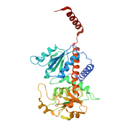
New Insight Into the Transcarbamylase Family: The Structure of Putrescine Transcarbamylase, a Key Catalyst for Fermentative Utilization of Agmatine
Polo, L.M., Gil-Ortiz, F., Cantin, A., Rubio, V.(2012) PLoS One 7: 31528
- PubMed: 22363663
- DOI: https://doi.org/10.1371/journal.pone.0031528
- Primary Citation of Related Structures:
4A8H, 4A8P, 4A8T - PubMed Abstract:
Transcarbamylases reversibly transfer a carbamyl group from carbamylphosphate (CP) to an amine. Although aspartate transcarbamylase and ornithine transcarbamylase (OTC) are well characterized, little was known about putrescine transcarbamylase (PTC), the enzyme that generates CP for ATP production in the fermentative catabolism of agmatine. We demonstrate that PTC (from Enterococcus faecalis), in addition to using putrescine, can utilize L-ornithine as a poor substrate. Crystal structures at 2.5 Å and 2.0 Å resolutions of PTC bound to its respective bisubstrate analog inhibitors for putrescine and ornithine use, N-(phosphonoacetyl)-putrescine and δ-N-(phosphonoacetyl)-L-ornithine, shed light on PTC preference for putrescine. Except for a highly prominent C-terminal helix that projects away and embraces an adjacent subunit, PTC closely resembles OTCs, suggesting recent divergence of the two enzymes. Since differences between the respective 230 and SMG loops of PTC and OTC appeared to account for the differential preference of these enzymes for putrescine and ornithine, we engineered the 230-loop of PTC to make it to resemble the SMG loop of OTCs, increasing the activity with ornithine and greatly decreasing the activity with putrescine. We also examined the role of the C-terminal helix that appears a constant and exclusive PTC trait. The enzyme lacking this helix remained active but the PTC trimer stability appeared decreased, since some of the enzyme eluted as monomers from a gel filtration column. In addition, truncated PTC tended to aggregate to hexamers, as shown both chromatographically and by X-ray crystallography. Therefore, the extra C-terminal helix plays a dual role: it stabilizes the PTC trimer and, by shielding helix 1 of an adjacent subunit, it prevents the supratrimeric oligomerizations of obscure significance observed with some OTCs. Guided by the structural data we identify signature traits that permit easy and unambiguous annotation of PTC sequences.
- Instituto de Biomedicina de Valencia and Centro de Investigación Biomédica en Red de Enfermedades Raras, Valencia, Spain.
Organizational Affiliation:


