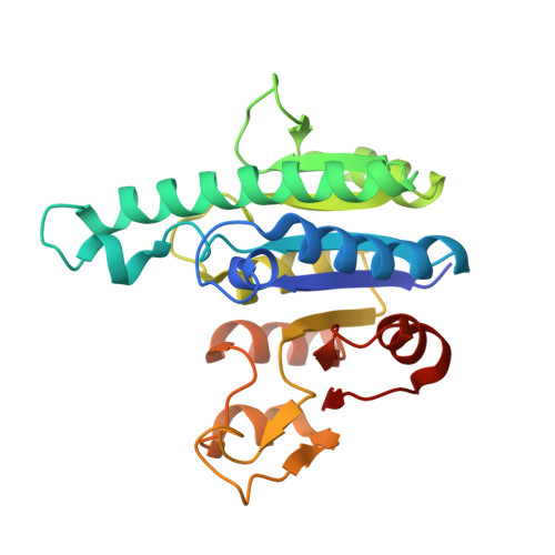
4A7X

Crystal structure of uridylate kinase from Helicobacter pylori


Experimental Data Snapshot

  • Method: X-RAY DIFFRACTION
  • Resolution: 2.49 Å
  • R-Value Free: 0.276 
  • R-Value Work: 0.213 
  • R-Value Observed: 0.216 

Starting Model: experimental
View more details

wwPDB Validation   3D Report Full Report


Ligand Structure Quality Assessment 


This is version 1.2 of the entry. See complete history


Literature

Structures of Helicobacter Pylori Uridylate Kinase: Insight Into Release of the Product Udp

Chu, C.H.Chen, P.C.Liu, M.H.Li, Y.C.Hsiao, C.D.Sun, Y.J.

(2012) Acta Crystallogr D Biol Crystallogr 68: 773

  • DOI: https://doi.org/10.1107/S0907444912011407
  • Primary Citation of Related Structures:  
    4A7W, 4A7X

  • PubMed Abstract: 

    Uridylate kinase (UMPK; EC 2.7.4.22) transfers the γ-phosphate of ATP to UMP, forming UDP. It is allosterically regulated by GTP. Structures of Helicobacter pylori UMPK (HpUMPK) complexed with GTP (HpUMPK-GTP) and with UDP (HpUMPK-UDP) were determined at 1.8 and 2.5 Å resolution, respectively. As expected, HpUMPK-GTP forms a hexamer with six GTP molecules at its centre. Interactions between HpUMPK and GTP are made by the β3 strand of the sheet, loop β3α4 and the α4 helix. In HpUMPK-UDP, the hexameric symmetry typical of UMPKs is absent. Only four of the HpUMPK molecules bind UDP; the other two HpUMPK molecules are in the UDP-free state. The asymmetric hexamer of HpUMPK-UDP, which has an exposed dimer interface, may assist in UDP release. Furthermore, the flexibility of the α2 helix, which interacts with UDP, is found to increase when UDP is absent in HpUMPK-UDP. In HpUMPK-GTP, the α2 helix is too flexible to be observed. This suggests that GTP binding may affect the conformation of the α2 helix, thereby promoting UDP release.


  • Organizational Affiliation

    Institute of Bioinformatics and Structural Biology, National Tsing Hua University, Hsinchu 300, Taiwan.


Macromolecules
Find similar proteins by:  (by identity cutoff)  |  3D Structure
Entity ID: 1
MoleculeChains Sequence LengthOrganismDetailsImage
URIDYLATE KINASE
A, B, C, D, E
A, B, C, D, E, F
240Helicobacter pylori 26695Mutation(s): 0 
EC: 2.7.4.22
UniProt
Find proteins for P56106 (Helicobacter pylori (strain ATCC 700392 / 26695))
Explore P56106 
Go to UniProtKB:  P56106
Entity Groups  
Sequence Clusters30% Identity50% Identity70% Identity90% Identity95% Identity100% Identity
UniProt GroupP56106
Sequence Annotations
Expand
  • Reference Sequence
Experimental Data & Validation

Experimental Data

  • Method: X-RAY DIFFRACTION
  • Resolution: 2.49 Å
  • R-Value Free: 0.276 
  • R-Value Work: 0.213 
  • R-Value Observed: 0.216 
  • Space Group: C 1 2 1
Unit Cell:
Length ( Å )Angle ( ˚ )
a = 147.136α = 90
b = 127.627β = 91.46
c = 93.181γ = 90
Software Package:
Software NamePurpose
REFMACrefinement
HKL-2000data reduction
SCALEPACKdata scaling
MOLREPphasing

Structure Validation

View Full Validation Report


Ligand Structure Quality Assessment 


Entry History 

Deposition Data

Revision History  (Full details and data files)

  • Version 1.0: 2012-06-27
    Type: Initial release
  • Version 1.1: 2012-07-11
    Changes: Other
  • Version 1.2: 2023-12-20
    Changes: Data collection, Database references, Derived calculations, Other, Refinement description