


The NMR Structure of Protoporphyrin-Ix Bound Murine P22Hbp
Goodfellow, B.J., Dias, J.S., Macedo, A.L., Ferreira, G.C., Peterson, F.C., Volkman, B.F., Duarte, I.C.N.To be published.
Experimental Data Snapshot
wwPDB Validation 3D Report Full Report

Entity ID: 1 | |||||
|---|---|---|---|---|---|
| Molecule | Chains | Sequence Length | Organism | Details | Image |
| HEME-BINDING PROTEIN 1 | 195 | Mus musculus | Mutation(s): 0 | 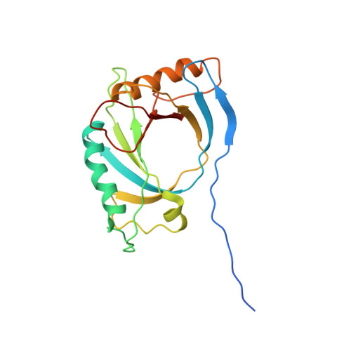 | |
UniProt | |||||
Entity Groups | |||||
| Sequence Clusters | 30% Identity50% Identity70% Identity90% Identity95% Identity100% Identity | ||||
| UniProt Group | Q9R257 | ||||
Sequence AnnotationsExpand | |||||
Reference Sequence | |||||


