 Download Mendeley
Crystal structure of the de novo designed miniprotein binder against PDL1, Gpx41 at pH 5.
Barik, T., Kumar, A., Gaikwad, S.S., Makde, R.D.To be published.
Experimental Data Snapshot
Starting Model: in silico
View more details
wwPDB Validation 3D Report Full Report

Entity ID: 1 | |||||
|---|---|---|---|---|---|
| Molecule | Chains | Sequence Length | Organism | Details | Image |
| De novo designed miniprotein | 73 | synthetic construct | Mutation(s): 0 | 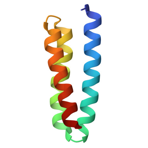 | |
Entity Groups | |||||
| Sequence Clusters | 30% Identity50% Identity70% Identity90% Identity95% Identity100% Identity | ||||
Sequence AnnotationsExpand | |||||
Reference Sequence | |||||
| Ligands 1 Unique | |||||
|---|---|---|---|---|---|
| ID | Chains | Name / Formula / InChI Key | 2D Diagram | 3D Interactions | |
| ACT Download:Ideal Coordinates CCD File | C [auth A] D [auth A] E [auth A] F [auth B] G [auth B] | ACETATE ION C2 H3 O2 QTBSBXVTEAMEQO-UHFFFAOYSA-M | 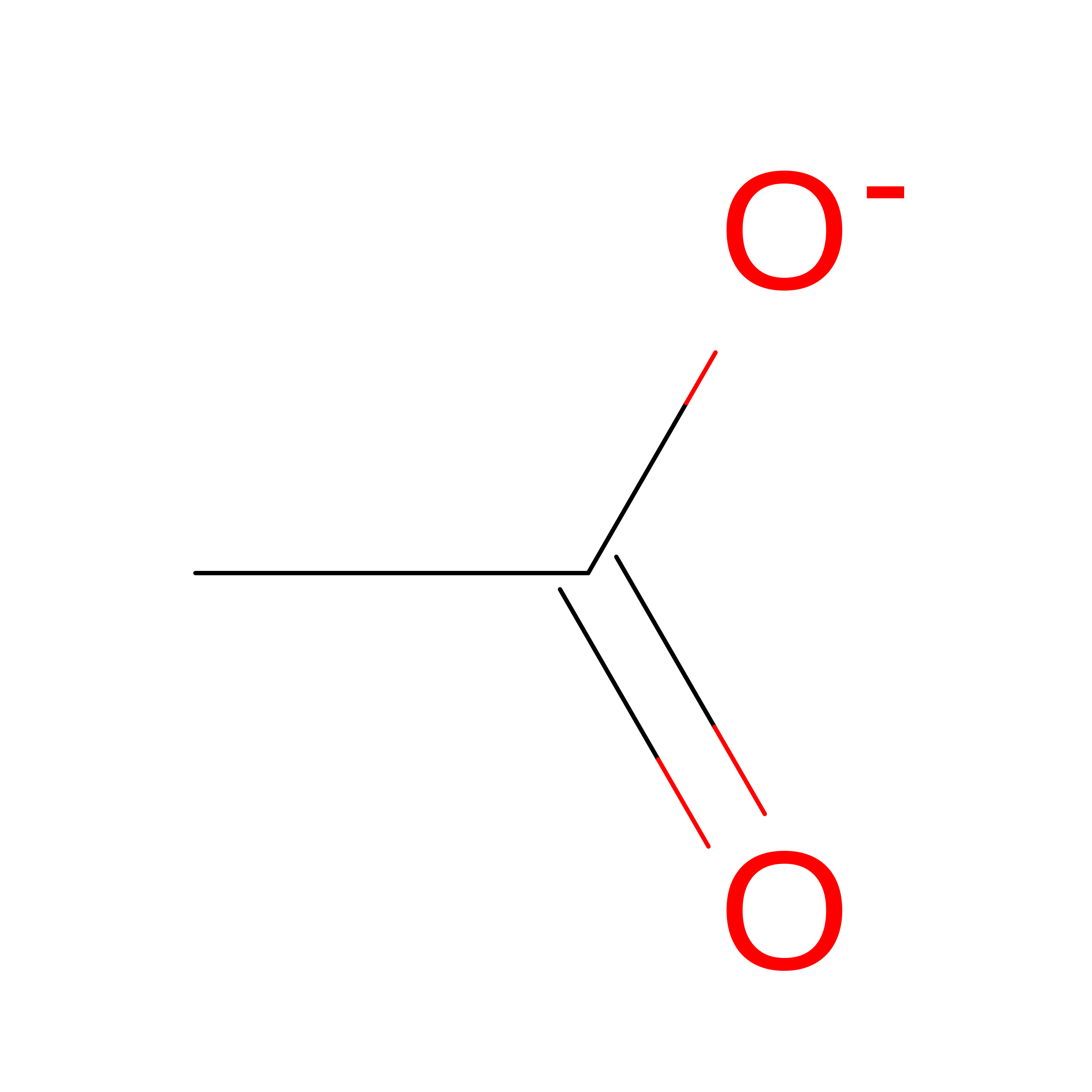 | ||
| Length ( Å ) | Angle ( ˚ ) |
|---|---|
| a = 44.818 | α = 90 |
| b = 44.818 | β = 90 |
| c = 157.528 | γ = 90 |
| Software Name | Purpose |
|---|---|
| XDS | data scaling |
| PHENIX | phasing |
| Coot | model building |
| PHENIX | refinement |
| XDS | data reduction |
| Funding Organization | Location | Grant Number |
|---|---|---|
| Other government | India | -- |
