456D | pdb_0000456d

MOLECULAR AND CRYSTAL STRUCTURE OF D(CGCGMO6AATCCGCG): THE WATSON-CRICK TYPE N6-METHOXYADENOSINE/CYTIDINE BASE-PAIRS IN B-DNA


Experimental Data Snapshot

  • Method: X-RAY DIFFRACTION
  • Resolution: 1.60 Å
  • R-Value Free: 
    0.231 (Depositor) 
  • R-Value Work: 
    0.196 (Depositor) 
  • R-Value Observed: 
    0.196 (Depositor) 

wwPDB Validation 3D Report Full Report

Validation slider image for 456D

This is version 1.3 of the entry. See complete history

Literature

Crystallographic studies on damaged DNAs. I. An N(6)-methoxyadenine residue forms a Watson-Crick pair with a cytosine residue in a B-DNA duplex.

Chatake, T.Ono, A.Ueno, Y.Matsuda, A.Takenaka, A.

(1999) J Mol Biology 294: 1215-1222

  • DOI: https://doi.org/10.1006/jmbi.1999.3303
  • Primary Citation Related Structures: 
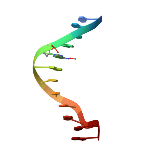
    456D

  • PubMed Abstract: 

    Oxyamines such as hydroxylamine and methoxylamine disturb DNA replication and act as potent mutagens, causing nucleotide transition from one purine to another or one pyrimidine to another. In order to investigate mismatch base-pairing in DNA damaged with oxyamines, a dodecamer with the sequence d(CGCGmo(6)AATCCGCG), where mo(6) A is 2'-deoxy-N(6)-methoxyadenosine, was synthesized and its crystal structure determined. No significant conformational changes are found between the present dodecamer and the original undamaged B-form dodecamer. Electron density maps clearly show that the mo(6)A residue forms a base-pair with a 2'-deoxycytidine residue through hydrogen bonds similar to a Watson-Crick G.C base-pair. For these hydrogen bonds to be made, N(6)-methoxyadenine must chemically take the imino form. The methoxylation thus enables the adenine base to mimic a guanine base. As a result, misincorporation of 2'-deoxycytidine instead of thymidine, or 2'-deoxyadenosine instead of 2'-deoxyguanosine, can occur in DNA replication.


  • Organizational Affiliation
    • Graduate School of Bioscience and Biotechnology, Tokyo Institute of Technology, Nagatsuda, Midori-ku, Yokohama, 226-8501, Japan.

Macromolecule Content 

  • Total Structure Weight: 7.38 kDa 
  • Atom Count: 662 
  • Modeled Residue Count: 24 
  • Deposited Residue Count: 24 
  • Unique nucleic acid chains: 1

Macromolecules

Find similar nucleic acids by:  Sequence
Entity ID: 1
MoleculeChains LengthOrganismImage
DNA (5'-D(*CP*GP*CP*GP*(A47)P*AP*TP*CP*CP*GP*CP*G)-3')
A, B
12N/A
Sequence Annotations
Expand
Reference Sequence

Small Molecules

Ligands 1 Unique
IDChains Name / Formula / InChI Key2D Diagram3D Interactions
MG

Query on MG


Download:Ideal Coordinates CCD File
C [auth A]MAGNESIUM ION
Mg
JLVVSXFLKOJNIY-UHFFFAOYSA-N

Experimental Data & Validation

Experimental Data

  • Method: X-RAY DIFFRACTION
  • Resolution: 1.60 Å
  • R-Value Free:  0.231 (Depositor) 
  • R-Value Work:  0.196 (Depositor) 
  • R-Value Observed: 0.196 (Depositor) 
Space Group: P 21 21 21
Unit Cell:
Length ( Å )Angle ( ˚ )
a = 25.1α = 90
b = 39.9β = 90
c = 65.8γ = 90
Software Package:
Software NamePurpose
AMoREphasing
X-PLORrefinement
DENZOdata reduction
CCP4data scaling

Structure Validation

View Full Validation Report


Entry History 

Deposition Data

Revision History  (Full details and data files)

  • Version 1.0: 2000-01-01
    Type: Initial release
  • Version 1.1: 2008-04-26
    Changes: Version format compliance
  • Version 1.2: 2011-07-13
    Changes: Version format compliance
  • Version 1.3: 2023-12-27
    Changes: Data collection, Database references, Derived calculations