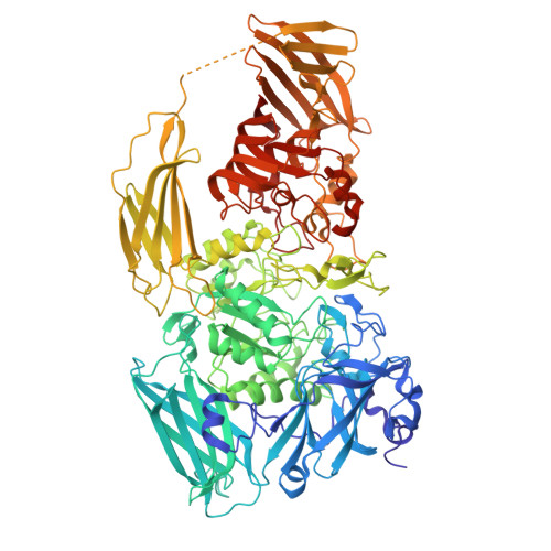
43LS | pdb_000043ls

Beta-galactosidase (without inhibitors) (E. coli)


Experimental Data Snapshot

  • Method: ELECTRON MICROSCOPY
  • Resolution: 1.36 Å
  • Aggregation State: PARTICLE 
  • Reconstruction Method: SINGLE PARTICLE 

wwPDB Validation 3D Report Full Report

Validation slider image for 43LS

This is version 1.0 of the entry. See complete history

Literature

Beta-galactosidase at 1.36 Angstrom

Burton-Smith, R.N.

To be published.

Macromolecule Content 

  • Total Structure Weight: 463.93 kDa 
  • Atom Count: 33,920 
  • Modeled Residue Count: 4,044 
  • Deposited Residue Count: 4,076 
  • Unique protein chains: 1

Macromolecules

Find similar proteins by:|  3D Structure
Entity ID: 1
MoleculeChains  Sequence LengthOrganismDetailsImage
Beta-galactosidase
A, B, C, D
1,019Escherichia coliMutation(s): 0 
EC: 3.2.1.23
UniProt
Find proteins for Q8VNN2 (Escherichia coli)
Explore Q8VNN2 
Go to UniProtKB:  Q8VNN2
Entity Groups
Sequence Clusters30% Identity50% Identity70% Identity90% Identity95% Identity100% Identity
UniProt GroupQ8VNN2
Sequence Annotations
Expand
Reference Sequence

Experimental Data & Validation

Experimental Data

  • Method: ELECTRON MICROSCOPY
  • Resolution: 1.36 Å
  • Aggregation State: PARTICLE 
  • Reconstruction Method: SINGLE PARTICLE 
EM Software:
TaskSoftware PackageVersion
RECONSTRUCTIONcryoSPARC

Structure Validation

View Full Validation Report


Entry History 

& Funding Information

Deposition Data


Funding OrganizationLocationGrant Number
Not funded--

Revision History  (Full details and data files)

  • Version 1.0: 2026-07-29
    Type: Initial release