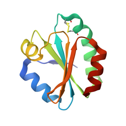
3ZZX

Crystallographic structure of thioredoxin from Litopenaeus vannamei


Experimental Data Snapshot

  • Method: X-RAY DIFFRACTION
  • Resolution: 1.88 Å
  • R-Value Free: 0.231 
  • R-Value Work: 0.182 
  • R-Value Observed: 0.185 

Starting Model: experimental
View more details

wwPDB Validation   3D Report Full Report


Ligand Structure Quality Assessment 


This is version 1.4 of the entry. See complete history


Literature

Expression, Purification, Crystallization and X-Ray Crystallographic Studies of Different Redox States of the Active Site of Thioredoxin 1 from the Whiteleg Shrimp Litopenaeus Vannamei

Campos-Acevedo, A.A.Garcia-Orozco, K.D.Sotelo-Mundo, R.R.Rudino-Pinera, E.

(2013) Acta Crystallogr Sect F Struct Biol Cryst Commun 69: 488

  • DOI: https://doi.org/10.1107/S1744309113010622
  • Primary Citation of Related Structures:  
    3ZZX, 4AJ6, 4AJ7, 4AJ8

  • PubMed Abstract: 

    Thioredoxin (Trx) is a 12 kDa cellular redox protein that belongs to a family of small redox proteins which undergo reversible oxidation to produce a cystine disulfide bond through the transfer of reducing equivalents from the catalytic site cysteine residues (Cys32 and Cys35) to a disulfide substrate. In this study, crystals of thioredoxin 1 from the Pacific whiteleg shrimp Litopenaeus vannamei (LvTrx) were successfully obtained. One data set was collected from each of four crystals at 100 K and the three-dimensional structures of the catalytic cysteines in different redox states were determined: reduced and oxidized forms at 2.00 Å resolution using data collected at a synchrotron-radiation source and two partially reduced structures at 1.54 and 1.88 Å resolution using data collected using an in-house source. All of the crystals belonged to space group P3212, with unit-cell parameters a = 57.5 (4), b = 57.5 (4), c = 118.1 (8) Å. The asymmetric unit contains two subunits of LvTrx, with a Matthews coefficient (VM) of 2.31 Å(3) Da(-1) and a solvent content of 46%. Initial phases were determined by molecular replacement using the crystallographic model of Trx from Drosophila melanogaster as a template. In the present work, LvTrx was overexpressed in Escherichia coli, purified and crystallized. Structural analysis of the different redox states at the Trx active site highlights its reactivity and corroborates the existence of a dimer in the crystal. In the crystallographic structures the dimer is stabilized by several interactions, including a disulfide bridge between Cys73 of each LvTrx monomer, a hydrogen bond between the side chain of Asp60 of each monomer and several hydrophobic interactions, with a noncrystallographic twofold axis.


  • Organizational Affiliation

    Departamento de Medicina Molecular y Bioprocesos, Instituto de Biotecnología (IBT), Universidad Nacional Autónoma de México (UNAM), Avenida Universidad 2001, Colonia Chamilpa, 62210 Cuernavaca, Morelos, Mexico.


Macromolecules
Find similar proteins by:  (by identity cutoff)  |  3D Structure
Entity ID: 1
MoleculeChains Sequence LengthOrganismDetailsImage
THIOREDOXIN
A, B
105Penaeus vannameiMutation(s): 0 
EC: 1.8.1.9
UniProt
Find proteins for B1PWB9 (Penaeus vannamei)
Explore B1PWB9 
Go to UniProtKB:  B1PWB9
Entity Groups  
Sequence Clusters30% Identity50% Identity70% Identity90% Identity95% Identity100% Identity
UniProt GroupB1PWB9
Sequence Annotations
Expand
  • Reference Sequence
Small Molecules
Ligands 4 Unique
IDChains Name / Formula / InChI Key2D Diagram3D Interactions
DTU
Query on DTU

Download Ideal Coordinates CCD File 
C [auth A](2R,3S)-1,4-DIMERCAPTOBUTANE-2,3-DIOL
C4 H10 O2 S2
VHJLVAABSRFDPM-ZXZARUISSA-N
SO4
Query on SO4

Download Ideal Coordinates CCD File 
M [auth B]SULFATE ION
O4 S
QAOWNCQODCNURD-UHFFFAOYSA-L
GOL
Query on GOL

Download Ideal Coordinates CCD File 
I [auth A],
J [auth A],
K [auth A],
L [auth A]
GLYCEROL
C3 H8 O3
PEDCQBHIVMGVHV-UHFFFAOYSA-N
ACT
Query on ACT

Download Ideal Coordinates CCD File 
D [auth A]
E [auth A]
F [auth A]
G [auth A]
H [auth A]
D [auth A],
E [auth A],
F [auth A],
G [auth A],
H [auth A],
N [auth B],
O [auth B]
ACETATE ION
C2 H3 O2
QTBSBXVTEAMEQO-UHFFFAOYSA-M
Experimental Data & Validation

Experimental Data

  • Method: X-RAY DIFFRACTION
  • Resolution: 1.88 Å
  • R-Value Free: 0.231 
  • R-Value Work: 0.182 
  • R-Value Observed: 0.185 
  • Space Group: P 32 1 2
Unit Cell:
Length ( Å )Angle ( ˚ )
a = 57.776α = 90
b = 57.776β = 90
c = 118.094γ = 120
Software Package:
Software NamePurpose
PHENIXrefinement
XDSdata reduction
SCALAdata scaling
PHASERphasing

Structure Validation

View Full Validation Report


Ligand Structure Quality Assessment 


Entry History 

Deposition Data

Revision History  (Full details and data files)

  • Version 1.0: 2012-09-26
    Type: Initial release
  • Version 1.1: 2013-05-15
    Changes: Database references
  • Version 1.2: 2013-06-05
    Changes: Database references
  • Version 1.3: 2023-12-20
    Changes: Data collection, Database references, Derived calculations, Other, Refinement description
  • Version 1.4: 2024-11-06
    Changes: Structure summary