3ZZJ | pdb_00003zzj

Structure of an engineered aspartate aminotransferase


Experimental Data Snapshot

  • Method: X-RAY DIFFRACTION
  • Resolution: 2.50 Å
  • R-Value Free: 
    0.262 (Depositor), 0.259 (DCC) 
  • R-Value Work: 
    0.217 (Depositor), 0.214 (DCC) 
  • R-Value Observed: 
    0.219 (Depositor) 

Starting Model: experimental
View more details

wwPDB Validation 3D Report Full Report

Validation slider image for 3ZZJ

Ligand Structure Quality Assessment 


This is version 1.1 of the entry. See complete history

Literature

Structure and Mechanism of a Cysteine Sulfinate Desulfinase Engineered on the Aspartate Aminotransferase Scaffold.

Fernandez, F.J.De Vries, D.Pena-Soler, E.Coll, M.Christen, P.Gehring, H.Vega, M.C.

(2011) Biochim Biophys Acta 1824: 339

  • DOI: https://doi.org/10.1016/j.bbapap.2011.10.016
  • Primary Citation Related Structures: 
    3ZZJ, 3ZZK, 4A00

  • PubMed Abstract: 

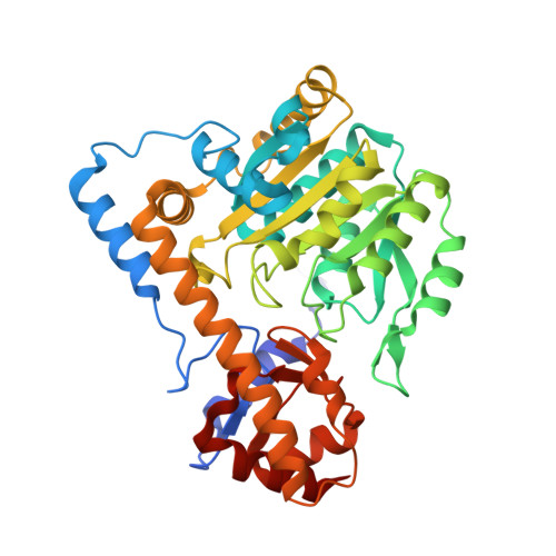
    The joint substitution of three active-site residues in Escherichia coli (L)-aspartate aminotransferase increases the ratio of l-cysteine sulfinate desulfinase to transaminase activity 10(5)-fold. This change in reaction specificity results from combining a tyrosine-shift double mutation (Y214Q/R280Y) with a non-conservative substitution of a substrate-binding residue (I33Q). Tyr214 hydrogen bonds with O3 of the cofactor and is close to Arg374 which binds the α-carboxylate group of the substrate; Arg280 interacts with the distal carboxylate group of the substrate; and Ile33 is part of the hydrophobic patch near the entrance to the active site, presumably participating in the domain closure essential for the transamination reaction. In the triple-mutant enzyme, k(cat)' for desulfination of l-cysteine sulfinate increased to 0.5s(-1) (from 0.05s(-1) in wild-type enzyme), whereas k(cat)' for transamination of the same substrate was reduced from 510s(-1) to 0.05s(-1). Similarly, k(cat)' for β-decarboxylation of l-aspartate increased from<0.0001s(-1) to 0.07s(-1), whereas k(cat)' for transamination was reduced from 530s(-1) to 0.13s(-1). l-Aspartate aminotransferase had thus been converted into an l-cysteine sulfinate desulfinase that catalyzes transamination and l-aspartate β-decarboxylation as side reactions. The X-ray structures of the engineered l-cysteine sulfinate desulfinase in its pyridoxal-5'-phosphate and pyridoxamine-5'-phosphate form or liganded with a covalent coenzyme-substrate adduct identified the subtle structural changes that suffice for generating desulfinase activity and concomitantly abolishing transaminase activity toward dicarboxylic amino acids. Apparently, the triple mutation impairs the domain closure thus favoring reprotonation of alternative acceptor sites in coenzyme-substrate intermediates by bulk water.


  • Organizational Affiliation
    • Centro de Investigaciones Biológicas, Consejo Superior de Investigaciones Científicas (Spanish National Research Council, CSIC), Madrid, Spain.

Macromolecule Content 

  • Total Structure Weight: 44.8 kDa 
  • Atom Count: 3,249 
  • Modeled Residue Count: 396 
  • Deposited Residue Count: 396 
  • Unique protein chains: 1

Macromolecules

Find similar proteins by:|  3D Structure
Entity ID: 1
MoleculeChains  Sequence LengthOrganismDetailsImage
ASPARTATE AMINOTRANSFERASE396Escherichia coliMutation(s): 3 
EC: 2.6.1.1
UniProt
Find proteins for P00509 (Escherichia coli (strain K12))
Explore P00509 
Go to UniProtKB:  P00509
Entity Groups
Sequence Clusters30% Identity50% Identity70% Identity90% Identity95% Identity100% Identity
UniProt GroupP00509
Sequence Annotations
Expand
Reference Sequence

Small Molecules

Ligands 5 Unique
IDChains Name / Formula / InChI Key2D Diagram3D Interactions
PLP

Query on PLP


Download:Ideal Coordinates CCD File
B [auth A]PYRIDOXAL-5'-PHOSPHATE
C8 H10 N O6 P
NGVDGCNFYWLIFO-UHFFFAOYSA-N
PEG

Query on PEG


Download:Ideal Coordinates CCD File
E [auth A],
F [auth A]
DI(HYDROXYETHYL)ETHER
C4 H10 O3
MTHSVFCYNBDYFN-UHFFFAOYSA-N
SO4

Query on SO4


Download:Ideal Coordinates CCD File
C [auth A],
D [auth A]
SULFATE ION
O4 S
QAOWNCQODCNURD-UHFFFAOYSA-L
GOL

Query on GOL


Download:Ideal Coordinates CCD File
H [auth A],
I [auth A],
J [auth A],
K [auth A],
L [auth A]
GLYCEROL
C3 H8 O3
PEDCQBHIVMGVHV-UHFFFAOYSA-N
BME

Query on BME


Download:Ideal Coordinates CCD File
G [auth A]BETA-MERCAPTOETHANOL
C2 H6 O S
DGVVWUTYPXICAM-UHFFFAOYSA-N

Experimental Data & Validation

Experimental Data

  • Method: X-RAY DIFFRACTION
  • Resolution: 2.50 Å
  • R-Value Free:  0.262 (Depositor), 0.259 (DCC) 
  • R-Value Work:  0.217 (Depositor), 0.214 (DCC) 
  • R-Value Observed: 0.219 (Depositor) 
Space Group: C 2 2 21
Unit Cell:
Length ( Å )Angle ( ˚ )
a = 85.139α = 90
b = 154.2β = 90
c = 79.931γ = 90
Software Package:
Software NamePurpose
REFMACrefinement
XDSdata reduction
SCALAdata scaling
AMoREphasing

Structure Validation

View Full Validation Report


Ligand Structure Quality Assessment 


Entry History 

Deposition Data

Revision History  (Full details and data files)

  • Version 1.0: 2011-12-28
    Type: Initial release
  • Version 1.1: 2023-12-20
    Changes: Data collection, Database references, Derived calculations, Other, Refinement description