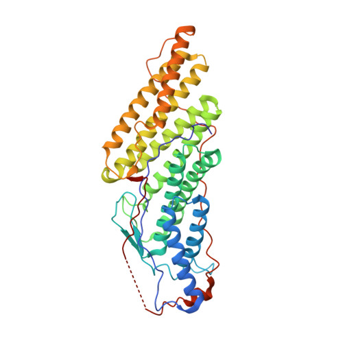
Structure of the Bro1 Domain Protein Brox and Functional Analyses of the Alix Bro1 Domain in HIV-1 Budding.
Zhai, Q., Landesman, M.B., Robinson, H., Sundquist, W.I., Hill, C.P.(2011) PLoS One 6: 27466
- PubMed: 22162750
- DOI: https://doi.org/10.1371/journal.pone.0027466
- Primary Citation of Related Structures:
3ZXP - PubMed Abstract:
Bro1 domains are elongated, banana-shaped domains that were first identified in the yeast ESCRT pathway protein, Bro1p. Humans express three Bro1 domain-containing proteins: ALIX, BROX, and HD-PTP, which function in association with the ESCRT pathway to help mediate intraluminal vesicle formation at multivesicular bodies, the abscission stage of cytokinesis, and/or enveloped virus budding. Human Bro1 domains share the ability to bind the CHMP4 subset of ESCRT-III proteins, associate with the HIV-1 NC(Gag) protein, and stimulate the budding of viral Gag proteins. The curved Bro1 domain structure has also been proposed to mediate membrane bending. To date, crystal structures have only been available for the related Bro1 domains from the Bro1p and ALIX proteins, and structures of additional family members should therefore aid in the identification of key structural and functional elements. We report the crystal structure of the human BROX protein, which comprises a single Bro1 domain. The Bro1 domains from BROX, Bro1p and ALIX adopt similar overall structures and share two common exposed hydrophobic surfaces. Surface 1 is located on the concave face and forms the CHMP4 binding site, whereas Surface 2 is located at the narrow end of the domain. The structures differ in that only ALIX has an extended loop that projects away from the convex face to expose the hydrophobic Phe105 side chain at its tip. Functional studies demonstrated that mutations in Surface 1, Surface 2, or Phe105 all impair the ability of ALIX to stimulate HIV-1 budding. Our studies reveal similarities in the overall folds and hydrophobic protein interaction sites of different Bro1 domains, and show that a unique extended loop contributes to the ability of ALIX to function in HIV-1 budding.
- Department of Biochemistry, University of Utah School of Medicine, Salt Lake City, Utah, United States of America.
Organizational Affiliation:


