3ZXD | pdb_00003zxd

wild-type lysenin


Experimental Data Snapshot

  • Method: X-RAY DIFFRACTION
  • Resolution: 3.30 Å
  • R-Value Free: 
    0.236 (Depositor), 0.259 (DCC) 
  • R-Value Work: 
    0.213 (Depositor), 0.236 (DCC) 
  • R-Value Observed: 
    0.214 (Depositor) 

Starting Model: experimental
View more details

wwPDB Validation 3D Report Full Report

Validation slider image for 3ZXD

This is version 1.2 of the entry. See complete history

Literature

Structures of Lysenin Reveal a Shared Evolutionary Origin for Pore-Forming Proteins and its Mode of Sphingomyelin Recognition.

De Colibus, L.Sonnen, A.F.P.Morris, K.J.Siebert, C.A.Abrusci, P.Plitzko, J.Hodnik, V.Leippe, M.Volpi, E.Anderluh, G.Gilbert, R.J.C.

(2012) Structure 20: 1498

  • DOI: https://doi.org/10.1016/j.str.2012.06.011
  • Primary Citation Related Structures: 
    3ZX7, 3ZXD, 3ZXG

  • PubMed Abstract: 

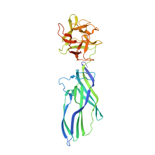
    Pore-forming proteins insert from solution into membranes to create lesions, undergoing a structural rearrangement often accompanied by oligomerization. Lysenin, a pore-forming toxin from the earthworm Eisenia fetida, specifically interacts with sphingomyelin (SM) and may confer innate immunity against parasites by attacking their membranes to form pores. SM has important roles in cell membranes and lysenin is a popular SM-labeling reagent. The structure of lysenin suggests common ancestry with other pore-forming proteins from a diverse set of eukaryotes and prokaryotes. The complex with SM shows the mode of its recognition by a protein in which both the phosphocholine headgroup and one acyl tail are specifically bound. Lipid interaction studies and assays using viable target cells confirm the functional reliance of lysenin on this form of SM recognition.


  • Organizational Affiliation
    • Division of Structural Biology, University of Oxford, Roosevelt Drive, Oxford OX3 7BN, UK.

Macromolecule Content 

  • Total Structure Weight: 141.05 kDa 
  • Atom Count: 9,376 
  • Modeled Residue Count: 1,166 
  • Deposited Residue Count: 1,236 
  • Unique protein chains: 1

Macromolecules

Find similar proteins by:|  3D Structure
Entity ID: 1
MoleculeChains  Sequence LengthOrganismDetailsImage
LYSENIN
A, B, C, D
309Eisenia fetidaMutation(s): 0 
UniProt
Find proteins for O18423 (Eisenia fetida)
Explore O18423 
Go to UniProtKB:  O18423
Entity Groups
Sequence Clusters30% Identity50% Identity70% Identity90% Identity95% Identity100% Identity
UniProt GroupO18423
Sequence Annotations
Expand
Reference Sequence

Small Molecules

Ligands 6 Unique
IDChains Name / Formula / InChI Key2D Diagram3D Interactions
MES

Query on MES


Download:Ideal Coordinates CCD File
BA [auth D],
N [auth A],
U [auth B]
2-(N-MORPHOLINO)-ETHANESULFONIC ACID
C6 H13 N O4 S
SXGZJKUKBWWHRA-UHFFFAOYSA-N
SO4

Query on SO4


Download:Ideal Coordinates CCD File
F [auth A]
H [auth A]
O [auth B]
P [auth B]
S [auth B]
F [auth A],
H [auth A],
O [auth B],
P [auth B],
S [auth B],
Y [auth D]
SULFATE ION
O4 S
QAOWNCQODCNURD-UHFFFAOYSA-L
GOL

Query on GOL


Download:Ideal Coordinates CCD File
E [auth A],
V [auth C]
GLYCEROL
C3 H8 O3
PEDCQBHIVMGVHV-UHFFFAOYSA-N
CL

Query on CL


Download:Ideal Coordinates CCD File
I [auth A]CHLORIDE ION
Cl
VEXZGXHMUGYJMC-UHFFFAOYSA-M
MG

Query on MG


Download:Ideal Coordinates CCD File
J [auth A]
Q [auth B]
R [auth B]
W [auth C]
X [auth C]
J [auth A],
Q [auth B],
R [auth B],
W [auth C],
X [auth C],
Z [auth D]
MAGNESIUM ION
Mg
JLVVSXFLKOJNIY-UHFFFAOYSA-N
NA

Query on NA


Download:Ideal Coordinates CCD File
AA [auth D]
G [auth A]
K [auth A]
L [auth A]
M [auth A]
AA [auth D],
G [auth A],
K [auth A],
L [auth A],
M [auth A],
T [auth B]
SODIUM ION
Na
FKNQFGJONOIPTF-UHFFFAOYSA-N

Experimental Data & Validation

Experimental Data

  • Method: X-RAY DIFFRACTION
  • Resolution: 3.30 Å
  • R-Value Free:  0.236 (Depositor), 0.259 (DCC) 
  • R-Value Work:  0.213 (Depositor), 0.236 (DCC) 
  • R-Value Observed: 0.214 (Depositor) 
Space Group: P 1
Unit Cell:
Length ( Å )Angle ( ˚ )
a = 58.91α = 98.88
b = 85.56β = 96.84
c = 108.81γ = 90.04
Software Package:
Software NamePurpose
BUSTERrefinement
XDSdata reduction
SCALAdata scaling
PHASERphasing

Structure Validation

View Full Validation Report


Entry History 

Revision History  (Full details and data files)

  • Version 1.0: 2012-09-19
    Type: Initial release
  • Version 1.1: 2012-10-03
    Changes: Database references
  • Version 1.2: 2023-12-20
    Changes: Data collection, Database references, Derived calculations, Other, Refinement description