3ZXA | pdb_00003zxa

Structure and Assembly of Turnip Crinkle Virus I. X-ray Crystallographic Structure Analysis at 3.2 A Resolution


Experimental Data Snapshot

  • Method: X-RAY DIFFRACTION
  • Resolution: 3.20 Å

Starting Model: other
View more details

wwPDB Validation 3D Report Full Report

Validation slider image for 3ZXA

This is version 1.5 of the entry. See complete history

Literature

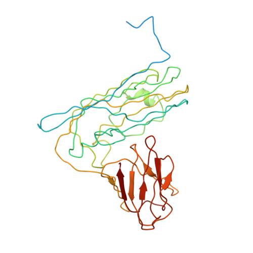
Structure and Assembly of Turnip Crinkle Virus. I. X-Ray Crystallographic Structure Analysis at 3.2 A Resolution.

Hogle, J.M.Maeda, A.Harrison, S.C.

(1986) J Mol Biology 191: 625

  • DOI: https://doi.org/10.1016/0022-2836(86)90450-x
  • Primary Citation Related Structures: 
    3ZXA

  • PubMed Abstract: 

    The structure of turnip crinkle virus has been determined at 3.2 A resolution, using the electron density of tomato bushy stunt virus as a starting point for phase refinement by non-crystallographic symmetry. The structures are very closely related, especially in the subunit arm and S domain, where only small insertions and deletions and small co-ordinate shifts relate one chain to another. The P domains, although quite similar in fold, are oriented somewhat differently with respect to the S domains. Understanding of the structure of turnip crinkle virus has been important for analyzing its assembly, as described in an accompanying paper.

Macromolecule Content 

  • Total Structure Weight: 37.76 kDa 
  • Atom Count: 2,237 
  • Modeled Residue Count: 295 
  • Deposited Residue Count: 347 
  • Unique protein chains: 1

Macromolecules

Find similar proteins by:|  3D Structure
Entity ID: 1
MoleculeChains  Sequence LengthOrganismDetailsImage
CAPSID PROTEINA [auth C]347Turnip crinkle virusMutation(s): 1 
UniProt
Find proteins for P06663 (Turnip crinkle virus)
Explore P06663 
Go to UniProtKB:  P06663
Entity Groups
Sequence Clusters30% Identity50% Identity70% Identity90% Identity95% Identity100% Identity
UniProt GroupP06663
Sequence Annotations
Expand
Reference Sequence

Experimental Data & Validation

Experimental Data

  • Method: X-RAY DIFFRACTION
  • Resolution: 3.20 Å
Space Group: I 2 2 2
Unit Cell:
Length ( Å )Angle ( ˚ )
a = 348.8α = 90
b = 379.1β = 90
c = 397.4γ = 90
Software Package:
Software NamePurpose
OPTRONICSdata reduction
DIGITALdata reduction
SCAN12data reduction
HARVARDdata scaling
COMBINEphasing
FFTphasing
BILDERrefinement

Structure Validation

View Full Validation Report


Entry History 

Deposition Data

Revision History  (Full details and data files)

  • Version 1.0: 2012-02-08
    Type: Initial release
  • Version 1.1: 2015-03-04
    Changes: Database references
  • Version 1.2: 2016-02-03
    Changes: Derived calculations
  • Version 1.3: 2017-06-28
    Changes: Data collection
  • Version 1.4: 2019-11-13
    Changes: Data collection, Database references
  • Version 1.5: 2024-05-01
    Changes: Data collection, Database references, Derived calculations, Refinement description