3ZV7 | pdb_00003zv7

Torpedo californica Acetylcholinesterase Inhibition by Bisnorcymserine


Experimental Data Snapshot

  • Method: X-RAY DIFFRACTION
  • Resolution: 2.26 Å
  • R-Value Free: 
    0.247 (Depositor), 0.238 (DCC) 
  • R-Value Work: 
    0.206 (Depositor), 0.201 (DCC) 
  • R-Value Observed: 
    0.206 (Depositor) 

Starting Model: experimental
View more details

wwPDB Validation 3D Report Full Report

Validation slider image for 3ZV7

Ligand Structure Quality Assessment 


This is version 2.2 of the entry. See complete history

Literature

Kinetics of Torpedo Californica Acetylcholinesterase Inhibition by Bisnorcymserine and Crystal Structure of the Complex with its Leaving Group.

Bartolucci, C.Stojan, J.Yu, Q.S.Greig, N.H.Lamba, D.

(2012) Biochem J 444: 269

  • DOI: https://doi.org/10.1042/BJ20111675
  • Primary Citation Related Structures: 
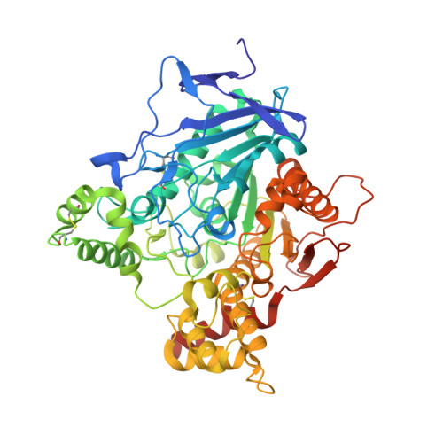
    3ZV7

  • PubMed Abstract: 

    Natural and synthetic carbamates act as pseudo-irreversible inhibitors of AChE (acetylcholinesterase) as well as BChE (butyrylcholinesterase), two enzymes involved in neuronal function as well as in the development and progression of AD (Alzheimer's disease). The AChE mode of action is characterized by a rapid carbamoylation of the active-site Ser(200) with release of a leaving group followed by a slow regeneration of enzyme action due to subsequent decarbamoylation. The experimental AD therapeutic bisnorcymserine, a synthetic carbamate, shows an interesting activity and selectivity for BChE, and its clinical development is currently being pursued. We undertook detailed kinetic studies on the activity of the carbamate bisnorcymserine with Tc (Torpedo californica) AChE and, on the basis of the results, crystallized the complex between TcAChE and bisnorcymserine. The X-ray crystal structure showed only the leaving group, bisnoreseroline, trapped at the bottom of the aromatic enzyme gorge. Specifically, bisnoreseroline interacts in a non-covalent way with Ser(200) and His(440), disrupting the existing interactions within the catalytic triad, and it stacks with Trp(84) at the bottom of the gorge, giving rise to an unprecedented hydrogen-bonding contact. These interactions point to a dominant reversible inhibition mechanism attributable to the leaving group, bisnoreseroline, as revealed by kinetic analysis.


  • Organizational Affiliation
    • Istituto di Cristallografia, Consiglio Nazionale delle Ricerche, Area della Ricerca di Roma, Monterotondo Scalo, Italy. cecilia.bartolucci@ic.cnr.it

Macromolecule Content 

  • Total Structure Weight: 63.15 kDa 
  • Atom Count: 4,669 
  • Modeled Residue Count: 535 
  • Deposited Residue Count: 543 
  • Unique protein chains: 1

Macromolecules

Find similar proteins by:|  3D Structure
Entity ID: 1
MoleculeChains  Sequence LengthOrganismDetailsImage
ACETYLCHOLINESTERASE543Tetronarce californicaMutation(s): 0 
EC: 3.1.1.7
UniProt
Find proteins for P04058 (Tetronarce californica)
Explore P04058 
Go to UniProtKB:  P04058
Entity Groups
Sequence Clusters30% Identity50% Identity70% Identity90% Identity95% Identity100% Identity
UniProt GroupP04058
Glycosylation
Glycosylation Sites: 2
Sequence Annotations
Expand
Reference Sequence

Oligosaccharides

Help  
Entity ID: 2
MoleculeChains Length2D Diagram GlycosylationD Interactions
2-acetamido-2-deoxy-beta-D-glucopyranose-(1-4)-2-acetamido-2-deoxy-beta-D-glucopyranose
B
2N-Glycosylation
Glycosylation Resources
GlyTouCan: G42666HT
GlyCosmos: G42666HT
GlyGen: G42666HT

Small Molecules

Ligands 7 Unique
IDChains Name / Formula / InChI Key2D Diagram3D Interactions
1PE

Query on 1PE


Download:Ideal Coordinates CCD File
C [auth A]PENTAETHYLENE GLYCOL
C10 H22 O6
JLFNLZLINWHATN-UHFFFAOYSA-N
NAG

Query on NAG


Download:Ideal Coordinates CCD File
H [auth A]2-acetamido-2-deoxy-beta-D-glucopyranose
C8 H15 N O6
OVRNDRQMDRJTHS-FMDGEEDCSA-N
MES

Query on MES


Download:Ideal Coordinates CCD File
G [auth A]2-(N-MORPHOLINO)-ETHANESULFONIC ACID
C6 H13 N O4 S
SXGZJKUKBWWHRA-UHFFFAOYSA-N
NHG

Query on NHG


Download:Ideal Coordinates CCD File
F [auth A]BIS-NORESEROLINE
C11 H14 N2 O
BLEHTFVVORLVPX-MNOVXSKESA-N
PEG

Query on PEG


Download:Ideal Coordinates CCD File
D [auth A]DI(HYDROXYETHYL)ETHER
C4 H10 O3
MTHSVFCYNBDYFN-UHFFFAOYSA-N
EDO

Query on EDO


Download:Ideal Coordinates CCD File
E [auth A]1,2-ETHANEDIOL
C2 H6 O2
LYCAIKOWRPUZTN-UHFFFAOYSA-N
CL

Query on CL


Download:Ideal Coordinates CCD File
I [auth A]
J [auth A]
K [auth A]
L [auth A]
M [auth A]
I [auth A],
J [auth A],
K [auth A],
L [auth A],
M [auth A],
N [auth A],
O [auth A],
P [auth A],
Q [auth A],
R [auth A],
S [auth A]
CHLORIDE ION
Cl
VEXZGXHMUGYJMC-UHFFFAOYSA-M

Experimental Data & Validation

Experimental Data

  • Method: X-RAY DIFFRACTION
  • Resolution: 2.26 Å
  • R-Value Free:  0.247 (Depositor), 0.238 (DCC) 
  • R-Value Work:  0.206 (Depositor), 0.201 (DCC) 
  • R-Value Observed: 0.206 (Depositor) 
Space Group: P 31 2 1
Unit Cell:
Length ( Å )Angle ( ˚ )
a = 111.008α = 90
b = 111.008β = 90
c = 137.387γ = 120
Software Package:
Software NamePurpose
CNSrefinement
DENZOdata reduction
SCALEPACKdata scaling
AMoREphasing

Structure Validation

View Full Validation Report


Ligand Structure Quality Assessment 


Entry History 

Deposition Data

Revision History  (Full details and data files)

  • Version 1.0: 2012-05-23
    Type: Initial release
  • Version 1.1: 2019-01-30
    Changes: Data collection, Experimental preparation, Other
  • Version 1.2: 2019-02-06
    Changes: Data collection, Experimental preparation
  • Version 2.0: 2020-07-29
    Type: Remediation
    Reason: Carbohydrate remediation
    Changes: Atomic model, Data collection, Derived calculations, Other, Structure summary
  • Version 2.1: 2023-12-20
    Changes: Data collection, Database references, Refinement description, Structure summary
  • Version 2.2: 2024-10-16
    Changes: Structure summary