3ZUK | pdb_00003zuk

CRYSTAL STRUCTURE OF MYCOBACTERIUM TUBERCULOSIS ZINC METALLOPROTEASE ZMP1 IN COMPLEX WITH INHIBITOR


Experimental Data Snapshot

  • Method: X-RAY DIFFRACTION
  • Resolution: 2.60 Å
  • R-Value Free: 
    0.250 (Depositor), 0.312 (DCC) 
  • R-Value Work: 
    0.175 (Depositor), 0.185 (DCC) 
  • R-Value Observed: 
    0.177 (Depositor) 

Starting Model: experimental
View more details

wwPDB Validation 3D Report Full Report

Validation slider image for 3ZUK

Ligand Structure Quality Assessment 


This is version 1.3 of the entry. See complete history

Literature

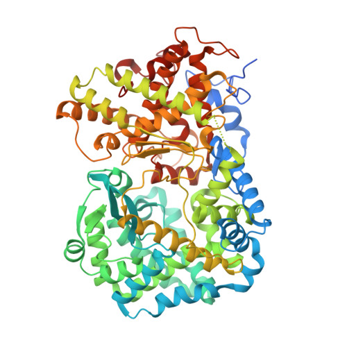
Crystal Structure of Mycobacterium Tuberculosis Zinc-Dependent Metalloprotease-1 (Zmp1), a Metalloprotease Involved in Pathogenicity.

Ferraris, D.M.Sbardella, D.Petrera, A.Marini, S.Amstutz, B.Coletta, M.Sander, P.Rizzi, M.

(2011) J Biological Chem 286: 32475

  • DOI: https://doi.org/10.1074/jbc.M111.271809
  • Primary Citation Related Structures: 
    3ZUK

  • PubMed Abstract: 

    Mycobacterium tuberculosis, the causative agent of tuberculosis, parasitizes host macrophages. The resistance of the tubercle bacilli to the macrophage hostile environment relates to their ability to impair phagosome maturation and its fusion with the lysosome, thus preventing the formation of the phago-lysosome and eventually arresting the process of phagocytosis. The M. tuberculosis zinc-dependent metalloprotease Zmp1 has been proposed to play a key role in the process of phagosome maturation inhibition and emerged as an important player in pathogenesis. Here, we report the crystal structure of wild-type Zmp1 at 2.6 Å resolution in complex with the generic zinc metalloprotease inhibitor phosphoramidon, which we demonstrated to inhibit the enzyme potently. Our data represent the first structural characterization of a bacterial member of the zinc-dependent M13 endopeptidase family and revealed a significant degree of conservation with eukaryotic enzymes. However, structural comparison of the Zmp1-phosphoramidon complex with homologous human proteins neprilysin and endothelin-converting enzyme-1 revealed unique features of the Zmp1 active site to be exploited for the rational design of specific inhibitors that may prove useful as a pharmacological tool for better understanding Zmp1 biological function.


  • Organizational Affiliation
    • DISCAFF Department of Chemical, Food, Pharmaceutical and Pharmacological Sciences, University of Piemonte Orientale A Avogadro, 28100 Novara, Italy.

Macromolecule Content 

  • Total Structure Weight: 164.29 kDa 
  • Atom Count: 11,220 
  • Modeled Residue Count: 1,314 
  • Deposited Residue Count: 1,398 
  • Unique protein chains: 1

Macromolecules

Find similar proteins by:|  3D Structure
Entity ID: 1
MoleculeChains  Sequence LengthOrganismDetailsImage
ENDOPEPTIDASE, PEPTIDASE FAMILY M13
A, B
699Mycobacterium tuberculosis H37RvMutation(s): 0 
EC: 3.4.24
UniProt
Find proteins for O53649 (Mycobacterium tuberculosis (strain CDC 1551 / Oshkosh))
Explore O53649 
Go to UniProtKB:  O53649
Entity Groups
Sequence Clusters30% Identity50% Identity70% Identity90% Identity95% Identity100% Identity
UniProt GroupO53649
Sequence Annotations
Expand
Reference Sequence

Small Molecules

Ligands 10 Unique
IDChains Name / Formula / InChI Key2D Diagram3D Interactions
RDF

Query on RDF


Download:Ideal Coordinates CCD File
D [auth A],
JA [auth B]
N-ALPHA-L-RHAMNOPYRANOSYLOXY(HYDROXYPHOSPHINYL)-L-LEUCYL-L-TRYPTOPHAN
C23 H34 N3 O10 P
ZPHBZEQOLSRPAK-XLCYBJAPSA-N
PG4

Query on PG4


Download:Ideal Coordinates CCD File
CB [auth B],
DB [auth B],
EB [auth B],
Z [auth A]
TETRAETHYLENE GLYCOL
C8 H18 O5
UWHCKJMYHZGTIT-UHFFFAOYSA-N
PGE

Query on PGE


Download:Ideal Coordinates CCD File
AB [auth B]
BB [auth B]
U [auth A]
V [auth A]
W [auth A]
AB [auth B],
BB [auth B],
U [auth A],
V [auth A],
W [auth A],
X [auth A],
Y [auth A],
ZA [auth B]
TRIETHYLENE GLYCOL
C6 H14 O4
ZIBGPFATKBEMQZ-UHFFFAOYSA-N
211

Query on 211


Download:Ideal Coordinates CCD File
E [auth A]2,2',2''-NITRILOTRIETHANOL
C6 H15 N O3
GSEJCLTVZPLZKY-UHFFFAOYSA-N
PEG

Query on PEG


Download:Ideal Coordinates CCD File
F [auth A]
G [auth A]
H [auth A]
I [auth A]
J [auth A]
F [auth A],
G [auth A],
H [auth A],
I [auth A],
J [auth A],
K [auth A],
KA [auth B],
L [auth A],
LA [auth B],
M [auth A],
MA [auth B],
N [auth A],
NA [auth B],
O [auth A],
OA [auth B],
P [auth A],
PA [auth B],
Q [auth A],
QA [auth B],
R [auth A],
RA [auth B],
S [auth A],
SA [auth B],
T [auth A],
TA [auth B],
UA [auth B],
VA [auth B],
WA [auth B],
XA [auth B],
YA [auth B]
DI(HYDROXYETHYL)ETHER
C4 H10 O3
MTHSVFCYNBDYFN-UHFFFAOYSA-N
SO4

Query on SO4


Download:Ideal Coordinates CCD File
FA [auth A]
GA [auth A]
NB [auth B]
OB [auth B]
PB [auth B]
FA [auth A],
GA [auth A],
NB [auth B],
OB [auth B],
PB [auth B],
QB [auth B]
SULFATE ION
O4 S
QAOWNCQODCNURD-UHFFFAOYSA-L
ZN

Query on ZN


Download:Ideal Coordinates CCD File
C [auth A],
IA [auth B]
ZINC ION
Zn
PTFCDOFLOPIGGS-UHFFFAOYSA-N
ACT

Query on ACT


Download:Ideal Coordinates CCD File
AA [auth A]
BA [auth A]
CA [auth A]
DA [auth A]
EA [auth A]
AA [auth A],
BA [auth A],
CA [auth A],
DA [auth A],
EA [auth A],
FB [auth B],
GB [auth B],
HB [auth B],
IB [auth B],
JB [auth B],
KB [auth B],
LB [auth B],
MB [auth B]
ACETATE ION
C2 H3 O2
QTBSBXVTEAMEQO-UHFFFAOYSA-M
CA

Query on CA


Download:Ideal Coordinates CCD File
SB [auth B]CALCIUM ION
Ca
BHPQYMZQTOCNFJ-UHFFFAOYSA-N
CL

Query on CL


Download:Ideal Coordinates CCD File
HA [auth A],
RB [auth B],
TB [auth B]
CHLORIDE ION
Cl
VEXZGXHMUGYJMC-UHFFFAOYSA-M

Biologically Interesting Molecules (External Reference) 

1 Unique

Experimental Data & Validation

Experimental Data

  • Method: X-RAY DIFFRACTION
  • Resolution: 2.60 Å
  • R-Value Free:  0.250 (Depositor), 0.312 (DCC) 
  • R-Value Work:  0.175 (Depositor), 0.185 (DCC) 
  • R-Value Observed: 0.177 (Depositor) 
Space Group: P 21 21 2
Unit Cell:
Length ( Å )Angle ( ˚ )
a = 128.29α = 90
b = 198.77β = 90
c = 63.27γ = 90
Software Package:
Software NamePurpose
REFMACrefinement
MOSFLMdata reduction
SCALAdata scaling
PHASERphasing

Structure Validation

View Full Validation Report


Ligand Structure Quality Assessment 


Entry History 

Deposition Data

Revision History  (Full details and data files)

  • Version 1.0: 2011-08-03
    Type: Initial release
  • Version 1.1: 2011-09-21
    Changes: Database references
  • Version 1.2: 2013-03-06
    Changes: Other
  • Version 1.3: 2023-12-20
    Changes: Data collection, Database references, Derived calculations, Other, Refinement description