3ZU8 | pdb_00003zu8

STRUCTURE OF CBM3B OF MAJOR SCAFFOLDIN SUBUNIT SCAA FROM ACETIVIBRIO CELLULOLYTICUS DETERMINED ON THE NIKEL ABSORPTION EDGE


Experimental Data Snapshot

  • Method: X-RAY DIFFRACTION
  • Resolution: 1.80 Å
  • R-Value Free: 
    0.182 (Depositor), 0.166 (DCC) 
  • R-Value Work: 
    0.154 (Depositor) 
  • R-Value Observed: 
    0.155 (Depositor) 

wwPDB Validation 3D Report Full Report

Validation slider image for 3ZU8

This is version 1.2 of the entry. See complete history

Literature

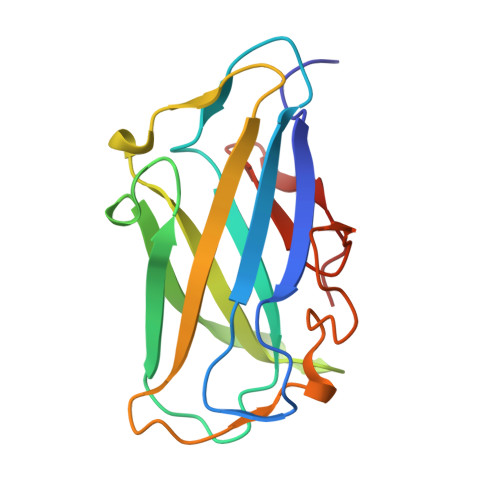
Structure of Cbm3B of the Major Scaffoldin Subunit Scaa from Acetivibrio Cellulolyticus

Yaniv, O.Halfon, Y.Shimon, L.J.W.Bayer, E.A.Lamed, R.Frolow, F.

(2012) Acta Crystallogr Sect F Struct Biol Cryst Commun 68: 8

  • DOI: https://doi.org/10.1107/S174430911104807X
  • Primary Citation Related Structures: 
    3ZQW, 3ZU8, 3ZUC

  • PubMed Abstract: 

    The carbohydrate-binding module (CBM) of the major scaffoldin subunit ScaA of the cellulosome of Acetivibrio cellulolyticus is classified as a family 3b CBM and binds strongly to cellulose. The CBM3b was overexpressed, purified and crystallized, and its three-dimensional structure was determined. The structure contained a nickel-binding site located at the N-terminal region in addition to a 'classical' CBM3b calcium-binding site. The structure was also determined independently by the SAD method using data collected at the Ni-absorption wavelength of 1.48395 Å and even at a wavelength of 0.97625 Å in a favourable case. The new scaffoldin-borne CBM3 structure reported here provides clear evidence for the proposition that a family 3b CBM may be accommodated in scaffoldin subunits and functions as the major substrate-binding entity of the cellulosome assembly.


  • Organizational Affiliation
    • Department of Molecular Microbiology and Biotechnology, Tel Aviv University, 69978 Tel Aviv, Israel.

Macromolecule Content 

  • Total Structure Weight: 17.19 kDa 
  • Atom Count: 1,467 
  • Modeled Residue Count: 153 
  • Deposited Residue Count: 153 
  • Unique protein chains: 1

Macromolecules

Find similar proteins by:|  3D Structure
Entity ID: 1
MoleculeChains  Sequence LengthOrganismDetailsImage
CELLULOSOMAL SCAFFOLDIN153Acetivibrio cellulolyticusMutation(s): 0 
EC: 3.2.1.4
UniProt
Find proteins for Q9RPL0 (Acetivibrio cellulolyticus)
Explore Q9RPL0 
Go to UniProtKB:  Q9RPL0
Entity Groups
Sequence Clusters30% Identity50% Identity70% Identity90% Identity95% Identity100% Identity
UniProt GroupQ9RPL0
Sequence Annotations
Expand
Reference Sequence

Experimental Data & Validation

Experimental Data

  • Method: X-RAY DIFFRACTION
  • Resolution: 1.80 Å
  • R-Value Free:  0.182 (Depositor), 0.166 (DCC) 
  • R-Value Work:  0.154 (Depositor) 
  • R-Value Observed: 0.155 (Depositor) 
Space Group: P 61 2 2
Unit Cell:
Length ( Å )Angle ( ˚ )
a = 52.263α = 90
b = 52.263β = 90
c = 192.938γ = 120
Software Package:
Software NamePurpose
PHENIXrefinement
MOSFLMdata reduction
SCALEPACKdata scaling
SHELXphasing

Structure Validation

View Full Validation Report


Entry History 

Deposition Data

Revision History  (Full details and data files)

  • Version 1.0: 2012-01-11
    Type: Initial release
  • Version 1.1: 2012-04-11
    Changes: Other
  • Version 1.2: 2024-05-08
    Changes: Data collection, Database references, Derived calculations, Other