3ZR1 | pdb_00003zr1

Crystal structure of human MTH1


Experimental Data Snapshot

  • Method: X-RAY DIFFRACTION
  • Resolution: 1.90 Å
  • R-Value Free: 
    0.245 (Depositor), 0.252 (DCC) 
  • R-Value Work: 
    0.191 (Depositor), 0.198 (DCC) 
  • R-Value Observed: 
    0.193 (Depositor) 

Starting Model: experimental
View more details

wwPDB Validation 3D Report Full Report

Validation slider image for 3ZR1

This is version 1.2 of the entry. See complete history

Literature

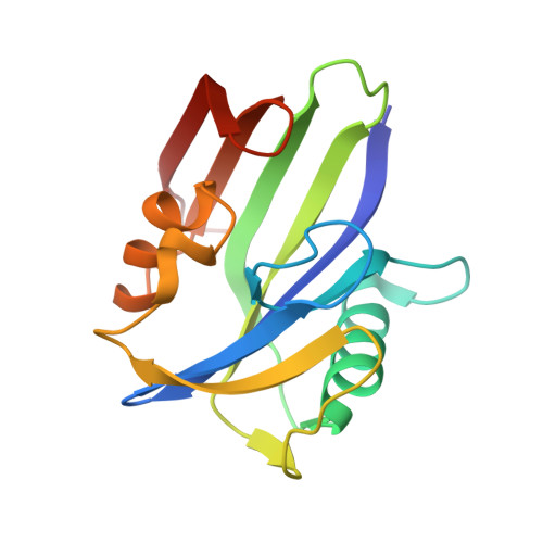
Crystal Structure of Human Mth1 and the 8-Oxo-Dgmp Product Complex.

Svensson, L.M.Jemth, A.Desroses, M.Loseva, O.Helleday, T.Hogbom, M.Stenmark, P.

(2011) FEBS Lett 585: 2617

  • DOI: https://doi.org/10.1016/j.febslet.2011.07.017
  • Primary Citation Related Structures: 
    3ZR0, 3ZR1

  • PubMed Abstract: 

    MTH1 hydrolyzes oxidized nucleotide triphosphates, thereby preventing them from being incorporated into DNA. We here present the structures of human MTH1 (1.9Å) and its complex with the product 8-oxo-dGMP (1.8Å). Unexpectedly MTH1 binds the nucleotide in the anti conformation with no direct interaction between the 8-oxo group and the protein. We suggest that the specificity depends on the stabilization of an enol tautomer of the 8-oxo form of dGTP. The binding of the product induces no major structural changes. The structures reveal the mode of nucleotide binding in MTH1 and provide the structural basis for inhibitor design.


  • Organizational Affiliation
    • Department of Biochemistry and Biophysics, Stockholm University, Stockholm, Sweden.

Macromolecule Content 

  • Total Structure Weight: 36.45 kDa 
  • Atom Count: 2,680 
  • Modeled Residue Count: 305 
  • Deposited Residue Count: 312 
  • Unique protein chains: 1

Macromolecules

Find similar proteins by:|  3D Structure
Entity ID: 1
MoleculeChains  Sequence LengthOrganismDetailsImage
7,8-DIHYDRO-8-OXOGUANINE TRIPHOSPHATASE
A, B
156Homo sapiensMutation(s): 0 
Gene Names: NUDT1
EC: 3.6.1 (PDB Primary Data), 3.6.1.56 (UniProt)
UniProt & NIH Common Fund Data Resources
Find proteins for P36639 (Homo sapiens)
Explore P36639 
Go to UniProtKB:  P36639
PHAROS:  P36639
GTEx:  ENSG00000106268 
Entity Groups
Sequence Clusters30% Identity50% Identity70% Identity90% Identity95% Identity100% Identity
UniProt GroupP36639
Sequence Annotations
Expand
Reference Sequence

Experimental Data & Validation

Experimental Data

  • Method: X-RAY DIFFRACTION
  • Resolution: 1.90 Å
  • R-Value Free:  0.245 (Depositor), 0.252 (DCC) 
  • R-Value Work:  0.191 (Depositor), 0.198 (DCC) 
  • R-Value Observed: 0.193 (Depositor) 
Space Group: P 21 21 21
Unit Cell:
Length ( Å )Angle ( ˚ )
a = 59.52α = 90
b = 67.08β = 90
c = 80.13γ = 90
Software Package:
Software NamePurpose
REFMACrefinement
MOSFLMdata reduction
SCALAdata scaling
BALBESphasing

Structure Validation

View Full Validation Report


Entry History 

Deposition Data

Revision History  (Full details and data files)

  • Version 1.0: 2011-07-27
    Type: Initial release
  • Version 1.1: 2011-08-24
    Changes: Database references
  • Version 1.2: 2023-12-20
    Changes: Data collection, Database references, Derived calculations, Other, Refinement description